
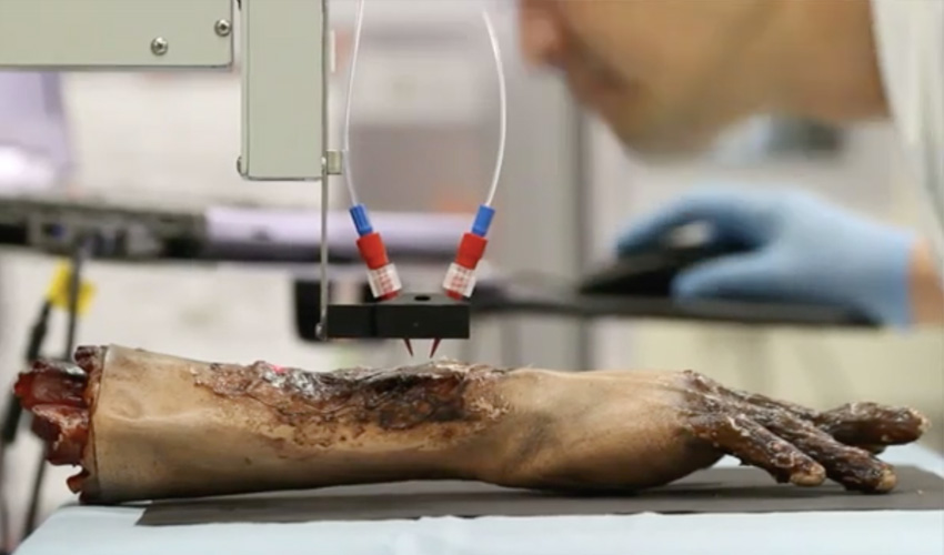
3D Bioprinting Solutions, a Russian bio-technical research laboratory, has announced plans to collaborate with scientists from the U.S. and Israel to deliver muscular tissue biomaterials to the International Space Station (ISS) in September as part of a microgravity 3D bioprinting experiment. The materials will be used with 3D Bioprinting Solutions’ Organ-Avt 3D bioprinter, which was […]
High-definition 3D printed organ models? German scientists think the answer is clear
Scientists at Ludwig Maximilians University in Munich have developed a new technique to make organs, and even whole organisms transparent – which aims to lead to high-fiedlity 3D printed models. The research team led by Dr. Ali Ertuerk developed the new technique, referred to as vDISCO, which uses a solvent to make living organs such […]
China: Complex GelMA-based Scaffolds Improved with the Addition of Nanoclay
Chinese scientists are delving further into successful bioprinting in ‘3D printing of complex GelMA-based scaffolds with nanoclay,’ exploring why photo-crosslinkable gelatin methacrylate (GelMA) has become so enticing for researchers attempting to engineer tissue. In a realm rife with obstacles, however, GelMA is no exception—constricted by viscosity issues and long cross-linking time.
The authors decided to bolster the ink further with nanoclay, in the hopes of being able to print stable, complex scaffolds. During this study, they evaluated windows for printability, issues with porosity and mechanical strength, and biocompatibility.
Obviously, without cell viability, there is no bioink and there are no spectacular innovations to write about. A wide range of hydrogels have been used successfully, with alginate commonly involved due to rapid crosslinking speed. Here, however, the researchers explain that alginate is not always conducive to attachment of cells or good function. Gelatin methacrylate (GelMA), however, is known to crosslink easily during light exposure. The researchers point out also that it maintains the biocompatibility of gelatin.
In attempting to overcome multiple issues with the use of GelMA, such as low viscosity and extensive time required for cross-linking, they examined the use of pre-crosslinking, post-crosslinking, in-situ crosslinking, and two-step crosslinking. Ultimately, the consensus was that all the methods were unsuitable, resulting in inferior stability. With the addition of nanoclay, however, the authors discovered that the ink had higher viscosity, and the GelMA scaffolds had better shape fidelity.
“After extrusion, the nanoclay rapidly converted to the gel state upon the release of shear stress, thereby forming stable hydrogel filament,” stated the authors. “Finally, the 3D structure was printed layer-by-layer by stacking the filament, and the GelMA within the filament was covalently crosslinked under UV light, resulting in a stable scaffold.”
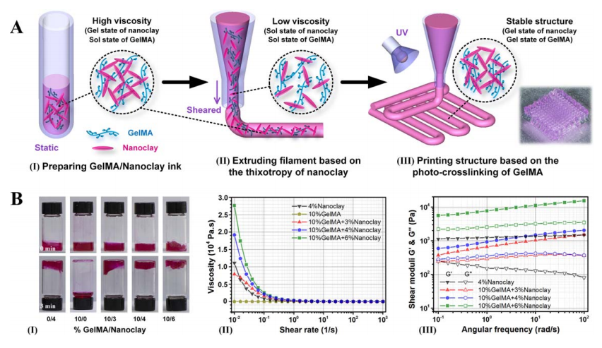
3D printing strategy of complex scaffolds with GelMA/Nanoclay ink. (A) Schematic illustration of printing scaffolds with GelMA/Nanoclay ink: (I) preparing GelMA/Nanoclay ink, (II) extruding filament based on the thixotropy property of nanoclay, and (III) printing structure based on the photo-crosslinking of GelMA. (B) Rheological properties of the GelMA/Nanoclay ink: (I) flow
behavior of 4% nanoclay, 10% GelMA, 10/3% GelMA/Nanoclay, 10/4% GelMA/Nanoclay, and 10/6% GelMA/Nanoclay, (II) the viscosity-shear rate, and (III) the shear moduli-angular frequency of the respective biomaterial inks.
They also found that nanoclay at higher levels resulted in less expansion due to more shear stress, meaning that nanoclay with higher concentration needed greater yield stress for deformation. In further discussion, the authors states that greater balance needs to researched for printing with GelMA/Nanoclay, and that so far, they surmise that if ‘cell-laden structures’ are to be directly 3D printed, they are forced to give up shape fidelity. Along with that, greater control is required of the following:
- Mechanical strength
- Degradation rate
- Tissue regeneration capacity
“Through systematic experiments that included rheological testing, printability analysis, property characterization, and biocompatibility characterization, we have answered several fundamental questions relating to this ink, including the formation mechanism for shear-thinning and rapid-gelling and the printability window for the fabrication of complex GelMA scaffolds, as well as showing that the addition of nanoclay improved the basic properties and had no effect on the excellent biological performance of the scaffolds,” concluded the researchers.
“Therefore, this method provides an easy way to fabricate complex GelMA-based scaffolds with good shape fidelity. It is very likely that this method will have versatile applications in the individualized therapy of tissue defects.”
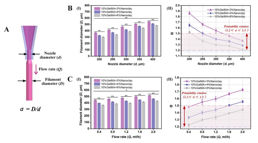
Printability analysis of GelMA/Nanoclay ink with regard to the extrusion process. (A) Schematic illustration of the expansion phenomenon and definition α = D/d.(B) Effect of the nozzle diameter on (I) the extruded filament diameter, and (II) α.
(C) Effect of the flow rate on (I) the extruded filament diameter, and (II) α.
The world of tissue engineering and bioprinting is rich in a variety of scaffolds, and while here we have learned more about the use of GelMA-based scaffolds, researchers around the world are constantly experimenting with new ways to sustain cells and make inks that are cell-laden. We have followed studies regarding transparent bioinks for fabricating corneas, making neural tissue, and 3D printing complex structures like alginate/gelatin hydrogels. What do you think of this news? Let us know your thoughts! Join the discussion of this and other 3D printing topics at 3DPrintBoard.com.
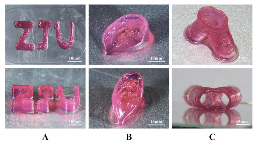
Photograph of the printed scaffolds with various shapes. (A) Abbreviation of Zhejiang University. (B) A bionic ear. (C)A branched vessel.
[Source / Images: ‘3D printing of complex GelMA-based scaffolds with nanoclay’]
Bioprinting 101 – Part 13 Imaging Technology

We talked about the way phantoms are created within bioprinting in our previous article. In this article, we will focus more on imaging technology that makes all of this possible. The medical field has been using imaging technology for quite some time for diagnosis, prognosis, as well as general healthcare applications. The scope of imaging technology has been widened with the interaction it is having with bioprinting, and we will look into imaging processes and how they affect this field.
Why is imaging so important for bioprinting you may ask? There is a large amount of data that is processed within an image. Images have resolution values that are determined by pixelation data. In order to process an image, one must digitize the image into a pixelated form. Pixelated images are the key to 3D models. In order to have a precise 3D model of a heart, we must have very high-resolution images within the 2D sphere. 2D images are stitched together to create a 3D transformed object. In terms of imaging technology, the better the original 2D images, a 3D object stitched from said images will have a higher quality in the overall look. When it comes to a 3D bioprinted object or phantom, we want to make sure that it has high fidelity and 3D image quality, thus the precision of different imaging technologies becomes very critical. We will talk about the advantages and disadvantages of each imaging technology in terms of bioprinting.
A computerized tomography (CT) scan combines a series of X-ray images taken from different angles around your body and uses computer processing to create cross-sectional images (slices) of the bones, blood vessels and soft tissues inside your body. CT scan images provide more-detailed information than plain X-rays do. A CT scan has many uses, but it’s particularly well-suited to quickly examine people who may have internal injuries from car accidents or other types of trauma. A CT scan can be used to visualize nearly all parts of the body and is used to diagnose disease or injury as well as to plan medical, surgical or radiation treatment. To bioprint with a layer-by-layer approach, tomographic reconstruction is done on the images. Tomographic reconstruction is a type of multidimensional inverse problem where the challenge is to yield an estimate of a specific system from a finite number of projections. The mathematical basis for tomographic imaging was laid down by Johann Radon. The system we are dealing with is the 3D dimensional object that is created through rotational geometric analysis on the multiple 2D images we have taken with a CT scanner. The now-2D images are then sent to the printer to be made. These cells are then mixed with a special liquefied material that provides oxygen and other nutrients to keep them alive. In some processes, the cells are encapsulated in cellular spheroids 500μm in diameter. This aggregation of cells does not require a scaffold, and are required for placing in the tubular-like tissue fusion for processes such as extrusion.
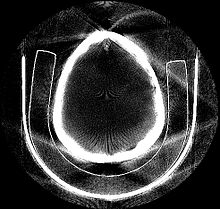 Tomographically Reconstructed Image
Magnetic resonance imaging (MRI) is a medical imaging technique used in radiology to form pictures of the anatomy and the physiological processes of the body in both health and disease. MRI scanners use strong magnetic fields, magnetic field gradients, and radio waves to generate images of the organs in the body. MRI does not involve X-rays or the use of ionizing radiation, which distinguishes it from CT or CAT scans and PET scans. Magnetic resonance imaging is a medical application of nuclear magnetic resonance (NMR). NMR can also be used for imaging in other NMR applications such as NMR spectroscopy.
Diagnostic ultrasound, also called sonography or diagnostic medical sonography, is an imaging method that uses high-frequency sound waves to produce images of structures within your body. The images can provide valuable information for diagnosing and treating a variety of diseases and conditions. Most ultrasound examinations are done using an ultrasound device outside your body, though some involve placing a device inside your body. Diagnostic ultrasound is a safe procedure that uses low-power sound waves. There are no known risks. Ultrasound is a valuable tool, but it has limitations. Sound doesn’t travel well through air or bone, so ultrasound isn’t effective at imaging body parts that have gas in them or are hidden by bone, such as the lungs or head. To view these areas, your doctor may order other imaging tests, such as CT or MRI scans or X-rays.
We need to explain specific software that helps to stitch images into 3D files. That software is called Mimics. Materialise Mimics is an image processing software for 3D design and modeling, developed by Materialise NV, a Belgian company specialized in additive manufacturing software and technology for medical, dental and additive manufacturing industries. Materialise Mimics is used to create 3D surface models from stacks of 2D image data. These 3D models can then be used for a variety of engineering applications. Mimics is an acronym for Materialise Interactive Medical Image Control System. It is developed in an ISO environment with CE and FDA 510k premarket clearance.
A major development within bioprinting is the development of imaging technology. Radiology and imaging allows for precision image quality that leads to overall higher resolution 3D models of structures of the body. At the previous RSNA conference in 2018, various 3D printing companies were in attendance showing the importance of Radiology and imaging technology when it comes to the future of 3D printing. This shows the way the industry is trending, and it will be interesting to see what can occur within the future. Precision is very important to the future of bioprinting. It will be cool to see how exact we can become with imaging and 3D model reconstruction.
Colombian Researchers Study Potential for SIS-Based Photocrosslinking in Bioinks

(a) Preparation of the 0.5% (w/v) riboflavin (RF) bioink and (b) its successful extrusion through a 21 G needle. (c) Filament formation during extrusion of the bioink through a 21 G needle. (d) Presumed photo-mediated crosslinking reaction thought to be occurring in the proposed bioinks.
Colombian researchers performed a recent study, outlined in ‘Formulation and Characterization of a SIS-Based Photocrosslinkable Bioink,’ explaining the possible value in crosslinking to create better materials for 3D printing cells. Here, they are using small intestinal submucosa (SIS) with photocrosslinked reactions to manipulate the gelation process, despite some expected challenges.
While the use of natural materials is always preferable, the researchers point out that they can also be difficult to work with due to lack of strength and stability. In the end that leads to inferior printability and further challenge.
“An avenue through which to overcome these issues is to mix them with synthetic polymers such as polyethylene glycol (PEG), polylactic acid (PLA), and polycaprolactone (PCL), which have demonstrated their ability to alter the mechanical response upon blending,” state the researchers. “Additionally, they have been proven able to shorten degradation rates, though at the expense of decreasing their biocompatibility.”
The authors also began to study decellularized extracellular matrices (dECMs) further, as they have the potential to copy the natural cellular environment. dECMs include the following proteins:
- Collagen
- Elastin
- Laminin
- Glycosaminoglycans
- Proteoglycans
- Growth factors
dECMs are not always stable though, and that presents challenges in bioprinting:
“Despite these obstacles, several research groups worldwide have attempted the development of bioinks based on dECMs,” state the researchers. “For inducing gelation, these studies have incorporated thermally-induced or photocrosslinking mechanisms, as well as a combination of the two.”
“Despite the crosslinking strategy implemented, the achieved mechanical stability has been observed to still not be sufficient, thereby requiring the use of synthetic materials as structural improvement supports.”
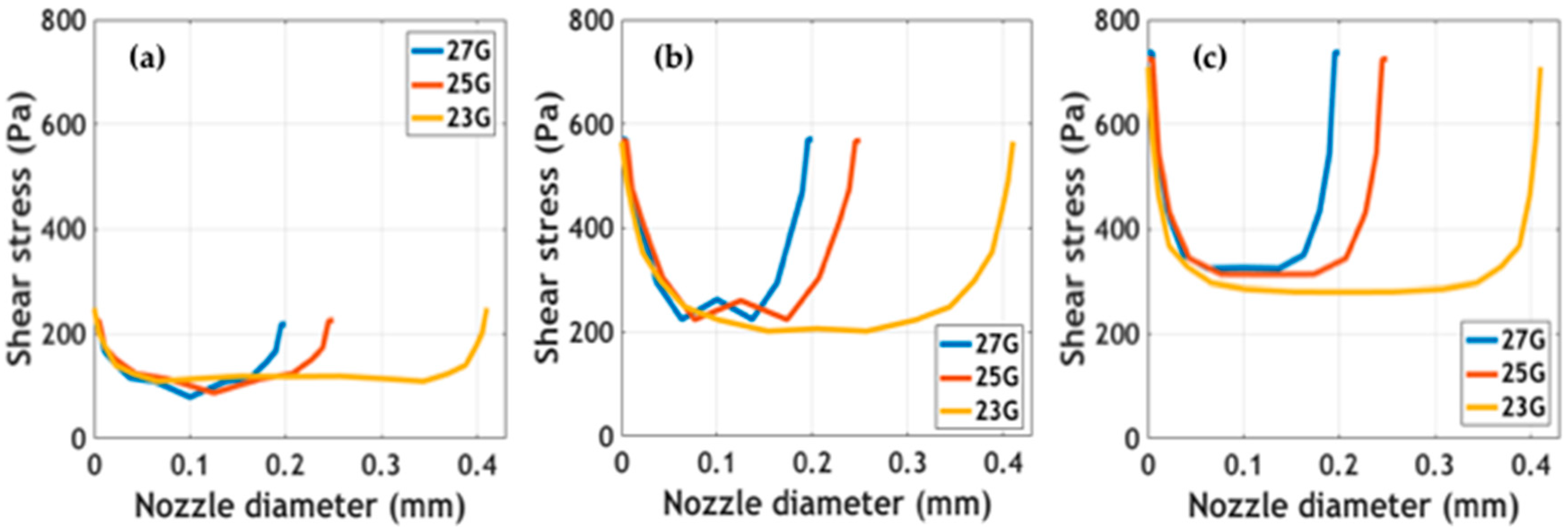
Shear stress profiles of the bioinks at different nozzle diameters and extrusion pressures: (a) 10 kPa, (b) 20 kPa, and (c) 60 kPa.
UV light has been used previously to increase the bioink stiffness in photocrosslinking, and for this study the authors experimented with the SIS dECM-based materials, using riboflavin (RF) as a photoinitiator. Visible light was used for the photocrosslinking. The research team created four different types of bioinks, with successful printability.
“Our experiments suggest that a successful extrusion can be accomplished while the pressure is maintained in the range 25–45 kPa,” stated the researchers.
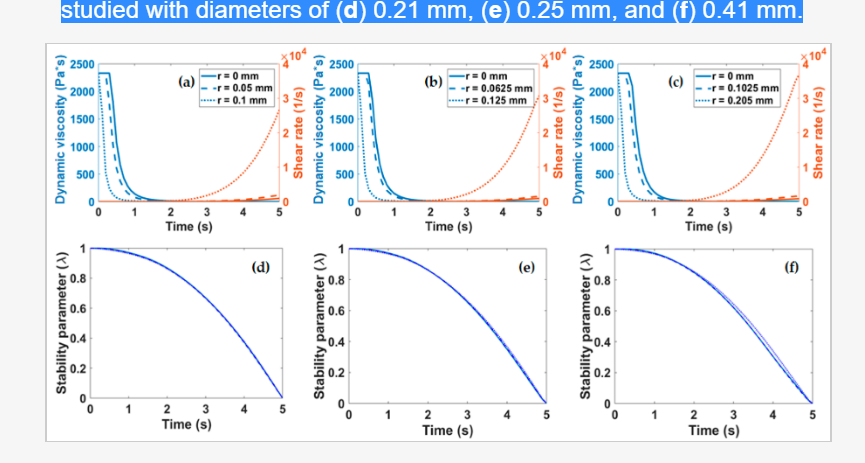
(a–c) Viscosity and shear rate as a function of time, measured at different points of the nozzle tip geometry (center, middle, and wall). Structural parameter for the three extrusion nozzles studied with diameters of (d) 0.21 mm, (e) 0.25 mm, and (f) 0.41 mm.
They also went on to state that these bioinks demonstrate strong mechanical properties that could ensure success in bioprinting endeavors—following in line with previous research studies where crosslinking resulted in excellent printability parameters, as well as offering better integrity in shape.
“Further in silico experiments allowed us to calculate a stability parameter that provided conceptual evidence for the aggregation of collagen in times as short as 5 s,” concluded the researchers. “Finally, rheology tests allowed us to recover power law parameters for CFD simulations that confirmed shear stress values low enough to maintain high cell viability levels.
“Future work will be focused on reformulating the bioink with the aid of synthetic polymers and/or thermal processing such that collagen fibers remain in an extended state and are readily accessible to the photoinitiated molecules.”
The study of materials in 3D printing has become a vast realm, and a necessary one for those dedicated to such progressive fabrication techniques. It is also a very serious area of study for scientists engaged in seeking out the best ways to grow tissue in the lab, with the potential for making serious impacts in medicine.
Researchers around the world are on an intense journey to perfect bioprinting, and eventually, reach the pinnacle of success in fabricating human organs. The challenge today, as tissue engineering results in a variety of different implants, is to keep cells alive to serve their function in bioprinting. This means seeking out the best bioprinted structures to build, bio-inks, printers, and techniques. Find out more about photocrosslinkable inks here. What do you think of this news? Let us know your thoughts! Join the discussion of this and other 3D printing topics at 3DPrintBoard.com.
[Source / Images: ‘Formulation and Characterization of a SIS-Based Photocrosslinkable Bioink’]
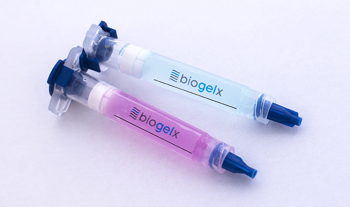
Biogelx Launching First Product Range of Synthetic Bioinks for Variety of 3D Printing Applications
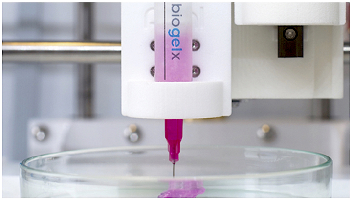
In 2013, a company called Biogelx was spun out from the University of Strathclyde in Glasgow, Scotland for the purposes of developing tunable, synthetic materials for use in 3D cell cultures and 3D bioprinting applications. Early on, the company worked on creating 3D cell culture scaffolds, which were in the form of synthetic peptide hydrogels so as to support cell growth by behaving as an extracellular matrix environment.
Bioinks are loaded up with cells in order to 3D print biological structures. Then, once the structure is printed, secondary crosslinking mechanisms help retain the shape’s structural fidelity. These bioprinting materials can facilitate cell adhesion, differentiation, and proliferation, as well as exhibit all the characteristics of an extracellular matrix environment. This is what makes it possible to create patient-specific human tissues in a laboratory setting, which is why good bioinks are very important.
![]() Biogelx’s hydrogel bioinks have unique physical and chemical tunability, which means they can successfully replicate specific tissue characteristics so cells can engage with, and experience, a 3D environment that’s pretty close to real life. Early on, the industry recognized how beneficial Biogelx’s hydrogel bioinks could be for researchers in the industry, as the materials claim to offer reproducibility, great printability, an easy crosslinking method, and viscosity control in one package. The company’s bioinks can provide a base modular material where the cells’ chemical and mechanical properties are able to be adapted.
Biogelx’s hydrogel bioinks have unique physical and chemical tunability, which means they can successfully replicate specific tissue characteristics so cells can engage with, and experience, a 3D environment that’s pretty close to real life. Early on, the industry recognized how beneficial Biogelx’s hydrogel bioinks could be for researchers in the industry, as the materials claim to offer reproducibility, great printability, an easy crosslinking method, and viscosity control in one package. The company’s bioinks can provide a base modular material where the cells’ chemical and mechanical properties are able to be adapted.
That’s why Biogelx is proud to launch its first product range of novel, synthetic bioinks: Biogelx-INKS. As the company says on its website, the future is synthetic.
“We are excited to announce the commercial availability of Biogelx
-INKs,” said Biogelx CEO, Mitch Scanlan. “Providing versatility and improving research outcomes are the key focuses for our product portfolio. We look forward to supporting researchers in their mission to develop realistic 3D disease modules, tissues, and organs for future pharmaceutical and medical applications.”
Developed with 3D printability at the forefront, Biogelx-INKs have been optimized for use in extrusion 3D printers, and they’re also versatile enough to work in 3D printing applications other than just bioprinting. The bioinks maintain the company’s core self-assembling peptide technology, and just like Biogelx’s hydrogel products, these new Biogelx-INKs form a nanofibrous network in order to mimic an extracellular matrix environment. This supports cell growth, proliferation, and signaling, but in addition, Biogelx-INKs were developed in such a way as to, as the company put it, “ensure the rheological properties are suitable for bioprinting applications.”
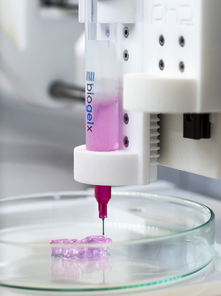 According to the Biogelx website, its new bioinks can be printed with good 3D fidelity, and don’t require any support, sacrificial, or curing inks. Biogelx-INKs also offer important shear-thinning behavior, which helps with cell viability, and controllable gelation, which is triggered by adding cell culture media and does not require the addition of reactive crosslinking reagents, UV curing, adjustments in pH, or extreme temperature. This feature also makes the material compatible with many different 3D bioprinters.
The company’s technology is based on a system of two peptides – a hydrophobic ‘gelator’ peptide (Fmoc-diphenylalanine) and a hydrophilic ‘surfactant’ (Fmoc-serine) – which self-assemble to form fibers in aqueous environments. These fibers have surface hydrophilic functionality, which is appropriate for cell adhesion.
Additional features of Biogelx-INKs include:
- Reproducible – these bioinks are totally synthetic and manufactured under strict quality control, which ensures batch-to-batch reproducibility and consistent prints.
- Tuneable – it’s possible to tailor the biomimetic functionality of the bioinks to specific cell types.
- Biocompatibility – composed of amino-acids, Biogelx-INKs are >95% water and produce a nanoscale structure which mimics the natural extracellular matrix environment.
- Material Biomimicry – this product range includes formulations that incorporate several biomimetic peptide sequences, which increases its biocompatibility with various cell types.
According to the Bioglex website, “With our core technology, our bioinks can be tailored to specific applications, meaning they have huge potential in a range of fields including cell research, toxicology, drug screening, and regenerative medicine.”
If you’re interested in the company’s new range of Biogelx-INKs, you can order the product, in one of three sizes, for £280.
Discuss this story and other 3D printing topics at 3DPrintBoard.com or share your thoughts in the Facebook comments below.
[Images: Biogelx]
Bioprinting 101 – Part 11, Tissue Engineering and Regenerative Medicine
I am glad to be running this series thus far. It seems that people are very interested with the subject matter, and I myself have learned even more as well. The series has discussed various technologies within bioprinting. We have also discussed a variety of bioprinting materials. We have barely even scratched the surface on topics we can talk about. In this article we are going to look into tissue engineering and regenerative medicine applications of 3D bioprinting.
Let us first define our terms of interest. Tissue engineering is the use of a combination of cells, engineering and materials methods, and suitable biochemical and physiochemical factors to improve or replace biological tissues. Tissue engineering involves the use of a tissue scaffolds for the formation of new viable tissue for a medical purpose. We have talked about the use of bioprinting scaffolds in order to create tissue layers for organs and other parts of the body. One must understand that as a bioengineer, most of the techniques used to create tissue is based on biomimicry. Biomimicry refers to the design and production of materials, structures, and systems that are modeled on biological entities and processes. We are limited in our scope due to the fact that biology is a complex system and it is difficult to mimic natural and living processes. 3D bioprinting is a tool that expedites biomimicry and allows us to create rapidly with synthetic resources.
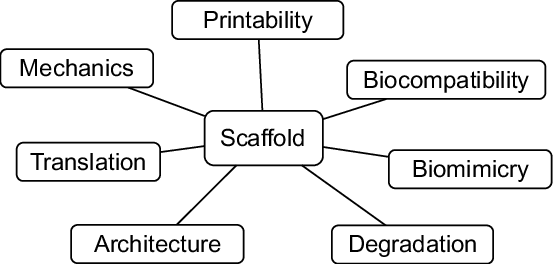
What is needed to create a scaffold for tissue engineering purposes
Regenerative medicine and tissue engineering go hand in hand. We are looking to build materials that may have innate regenerative properties within our bodies. This is a process that is essential to living organisms – the ability to repair oneself after damage or trauma. Regenerative medicine is a branch of translational research in tissue engineering and molecular biology which deals with the “process of replacing, engineering or regenerating human cells, tissues or organs to restore or establish normal function”. Extracellular matrix materials are commercially available and are used in reconstructive surgery, treatment of chronic wounds, and some orthopedic surgeries. This is the future of medical care in many ways. Our understanding of the ECM and bioprinting is evolving, and technology is continuously improving. The future of ECM biomaterials in tissue engineering and regenerative medicine applications is promising. Progress in decellularization techniques and optimization of recellularization strategies will improve various aspects of an ECM scaffold and its ability to be regenerative. These traits include biocompatibility, endothelialization, and functional anastomosis into the host vasculature.
Regenerative Medicine and 3D bioprinting
Endothelialization refers to the creation of endothelial tissue. Endothelial tissue refers to the tissue the layer of cells lining the inside of blood and lymph vessels, of the heart, and of some other closed cavities. Anastomosis a cross-connection between adjacent channels, tubes, fibers, or other parts of a network. These are major concerns when it comes to tissue engineering and regenerative medicine due to how we need to understand the need for biomimicry. We have to realize that it takes an absurd amount of precision to even build systems that replicate the inner structure of a vascular tissue layer. Then we have to think about how this will be self healing as well. This does not deter the development of this technology though as more people are intrigued by this subject matter on a daily basis.
3D bioprinting is an amazing step in the right direction, and there are companies, universities, and startups who are trying to be on the cutting edge of this field. The wealth of knowledge being created in this field is immense and frankly would intimidate people who are not aware of how vast the field is.
Now I believe the way for us to really delve deep within this series from now on is to start interviewing the leaders within this field. Due diligence will be done for the public to get more in depth understanding from industry experts. It is important to interview a variety of people within this field as no one necessarily is a specialist in this field. There seems to be a lot of cross pollination and multi skilled individuals. It makes for a wide variety of knowledge to be learned within the field of biology, synthetic biology, biomimicry, biophysics, chemistry, biochemistry, biomaterials, material science, biomedical fields, and a bunch of other fields I have not had a chance to mention. I personally believe that the revolution of bioprinting is a bit early. There is just so much development from different places. It is difficult to see the best methods at the moment. I will try to interview others to gain more insight and build a repertoire of knowledge for all readers.
This article is part of a series that wishes to make bioprinting more accessible. It starts with bioprinting 101, Hydrogels, 3D Industrial Bioprinters, Alginate, Bioinks, Pluronics, Applications, Gelatin, and Decellularized Extracellular Matrices.
Bioprinting 101 – Part 10, Disease Modeling
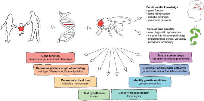
Disease modeling is an important field to understand in terms of human health for the present and the future. A disease model is an animal or cells displaying all or some of the pathological processes that are observed in the actual human or animal disease. Studying disease models aids understanding of how the disease develops and testing potential treatment approaches. We will take a look at how disease models are constructed currently and how bioprinting will help build disease models in the future.
One must know of some preliminary knowledge before we continue to discuss about bioprinting and disease modeling. Disease modeling utilizes different hosts to study underlying pathology of a disease. We can not experiment on a human due to moral and ethical grounds, so instead we typically use a mouse, rat, a pea plant, fish, nematodes, as well as fruit flies for our host site. We then inject said host with a particular disease and it allows us to study how the disease operates within that host. With disease models, we are able to use the information gathered from experimentation to figure out how we can prevent or reduce the spreading of different diseases within the human race.When animal models are employed in the study of human disease, they are frequently selected because of their similarity to humans in terms of genetics, anatomy, and physiology. Animal models are preferable for experimental disease research because of their unlimited supply and ease of manipulation. We can also utilize in vitro cell assays and inject these with different diseases. This allows us to study cell interaction as well.
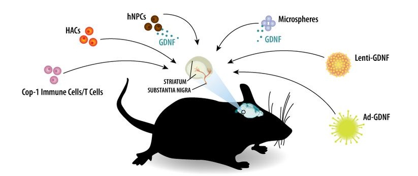
Rodent Model of Parkinson’s Disease
With the methodology roughly outlined, we will now explain how bioprinting is important for this field. Conventional two-dimensional (2D) in vitro assays and animal models have been unable to fully recapitulate the critical characteristics of human physiology. Physiology refers to how organisms, organ systems, organs, cells, and biomolecules carry out the chemical and physical functions that exist in a living system. Alternatively, three-dimensional (3D) tissue models are often developed in a low-throughput manner and lack crucial native-like architecture. The recent emergence of bioprinting technologies has enabled creating 3D tissue models that address the critical challenges of conventional in vitro assays through the development of custom bioinks and patient derived cells coupled with well-defined arrangements of biomaterials.
Bioprinting is usefull because we can derive materials from human cells to see better reactions without needing to mutate an animal host to become similar to the human genome. There are a variety of diseases that humans contrive but are not within different organisms and their respective genomes. This causes one to necessitate mutation within a host. Ideally one would stay away from mutation when studying disease as that may be a variable that causes imperfect interpretation of results and studies of pathology. Also it is important to consider that physiology of a human is very different from the physiology of a rat, mouse, a plant, and etc. With bioprinting we can actually create scaffolds that have physiological properties similar to human tissue. That allows for more accurate studies as well of interaction from a physiology and pathology framework.
There still lies some areas of concern in terms of bioprinting and disease modeling. It is important for a bioprinted material used in disease modeling to have the following properties:
- Ability to survive for longer time periods
- Allows for safe cell proliferation
- Allows for safe cell differentiation
In order to study a disease pathology and physiology one needs to be able to trust that the material we are using will be able to last and live long enough for useful data or information collected from experimentation. There does not seem to be a lot of research done currently on degradation times of bioprinted tissue scaffolds. This level of uncertainty causes disease modeling to shy away from bioprinting currently. It is important to have clinical viability, and uncertainty is not needed in clinical settings.
Cell proliferation refers to the process that result in an increase of the number of cells, and is defined by the balance between cell divisions and cell loss through cell death or differentiation. This can be further understood through mitosis. Mitosis refers to a type of cell division that results in two daughter cells each having the same number and kind of chromosomes as the parent nucleus, typical of ordinary tissue growth. It is difficult at this stage to induce such cell proliferation within bioprinted materials. There are various studies that show proliferation within bioprinted materials, but there is not enough work done to be able to safely control proliferation.
Cellular differentiation is the process where a cell changes from one cell type to another. Usually, the cell changes to a more specialized type. Differentiation occurs numerous times during the development of a multicellular organism as it changes from a simple zygote to a complex system of tissues and cell types. Cellular differentiation must be controllable. The ability to tune bioprinting properties as an approach to fabricate stem cell bearing scaffolds and to also harness the benefits of the cells multipotency is of considerable relevance to the field of biomaterials and bioengineering. Without a regimented method to control differentiation within a disease modeling context, various things may go awry within experimentation.
Proliferation and Differentiation
Overall, disease modelling is ripe for change. With bioprinting, the future is intriguing and will be more accurate in term of studying human pathology and physiology of diseases because of us having the ability to derive tissue from patient derived cells. There are still a number of items that need to be changed in order for bioprinting within the disease modelling context to be used more thoroughly, and researchers are attempting to fix those issues.
This article is part of a series that wishes to make bioprinting more accessible. It starts with bioprinting 101, Hydrogels, 3D Industrial Bioprinters, Alginate, Bioinks, Pluronics, Applications, Gelatin, Decellularized Extracellular Matrices, and Tissue Engineering & Regenerative Medicine.
Polish Company CD3D Opens Largest 3D Bioprinting Cluster in Europe
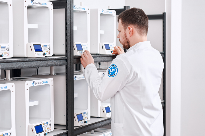
Centrum Druku 3D, or CD3D, is the largest online website devoted to 3D printing technology in Poland. Launched in 2013 with an online portal, the company’s operations are based on two important pillars: providing knowledge in the 3D printing field, and scientific-research and R&D activities in the medical and pharmaceutical sciences. In 2014, CD3D held Poland’s first 3D printing awards, and this week launched a new medical project – the largest 3D bioprinting center in Europe.
The Open 3D Bioprinting Cluster launched in Lodz at the Bionanopark, which is one of the country’s largest laboratory complexes and works on multiple science and research projects in the medicine and biotechnology fields, including computational chemistry, 3D printing, biochemistry, and medical implants. CD3D, under the CD3D Medical brand, is the creator of the cluster, and will be operating it together with the Laboratory of Molecular and Nanostructured Biophysics at the complex, which also includes an incubator and conference center. In addition to bioprinting, CD3D Medical also offers SLA, FDM, and DMP 3D printing technologies.
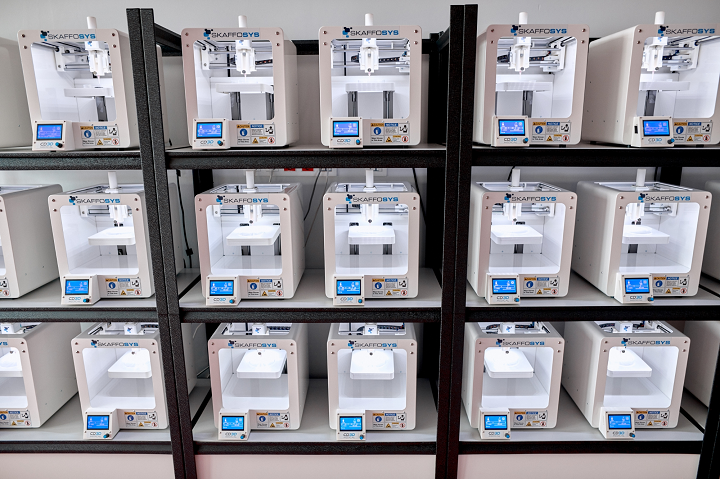 21 3D bioprinters, created by CD3D and called SKAFFOSYS for ‘scaffold systems’, make up the cluster, and according to Pawel Slusarczyk, a Project Director at CD3D, they are the first Polish bioprinters.
The system uses a 5 ml syringe as a printhead, and performs extrusion mechanically, as semi-liquid, gel, and hydrogel materials are applied to a laboratory pan that’s been affixed to a working table. The SKAFFOSYS Lite 3D bioprinter features a 170 x 125 x 80 mm build area, with a process accuracy of 0.2 mm, and can also complete bioplotting. As more challenges are created over time by bioprinting projects, CD3D will expand the SKAFFOSYS Lite by adding new functionalities and modules.
Due to the teamwork between the Bionanopark and CD3D Medical, scientists are able to use additive bioprinting to complete comprehensive research and development projects in the biomedical engineering field. Under the close supervision of CD3D specialists and scientists from the Laboratory of Molecular and Nanostructured Biophysics, laboratories at the Bionanopark can now successfully complete, according to the website, “biochemical, biological and molecular research at virtually any stage of the creation of three-dimensional structures.”

The reason the 3D Bioprinting Cluster is so important is due to its open nature. We use 3D bioprinted structures for a myriad of purposes, from growing biological material on printed scaffolds and creating composite materials to researching alternative food sources and creating, studying, and testing out new types of biocompatible materials. So the fact that this large, new cluster for 3D bioprinting is open means that other external entities can use its important resources to complete tasks such as commissioning a comprehensive scientific and research service.
The partners and customers of the new Open 3D Bioprinting Cluster in Poland can now rest assured that the comprehensive service will make it possible to outsource scientific research projects to all of the laboratories in the Bionanopark.
What do you think? Discuss this story and other 3D printing topics at 3DPrintBoard.com or share your thoughts in the Facebook comments below.
[Images: CD3D]
Biological Gradients Help Researchers Understand More in Bioprinting
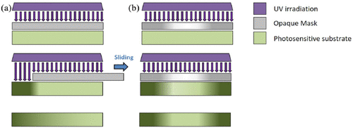
Selective irradiation method to induce linear or radial gradients on a photosensitive substrate. Ultraviolet (UV) irradiation is selectively filtered by a mask, which is either completely opaque (a) or has a designed distribution of opacity (b). The movement of the mask or the selective irradiation induces a controlled modification of the substrate, which results in a physical or chemical gradient.
Gradients present in structures today help us understand more about their properties, and this is critical in areas like bioprinting. Through replicating gradients we can begin to make accurate high resolution parts and scaffolds. Researchers explored these needs further in a recently published paper, ‘Engineering Biological Gradients.’ Tissue engineering is growing more and more of interest to researchers around the world as they continue to strive to 3D print human organs—a subject we have followed many times over the years as progress has been made in 3D printed kidneys, 3D printed livers, and much more.
In the meantime, many different devices and implants are being created in the realm of regenerative medicine—with the researchers here categorizing structures as either scaffolds or constructs (scaffolds combined with cells prior to implantation). As researchers seek to forge ahead with even fewer limits, they have begun measurement and analysis for clinical therapy and in engineering tissue for in-vitro screening, allowing for better feedback.
“The presence of a gradient confers to each point of the substrate a specific value of the varying quantity, allowing analysis of the effect of each variable over a specific phenomenon, such as cell adhesion, spreading, morphology, or differentiation,” stated the researchers. “This offers a great advantage to both interfaced tissue engineering and drug screening, as the effect of different variables on a phenomenon is analyzed in a single experimental set-up rather than in a series of experiments at different conditions.”
Gradients are basically variables used in a range of different measurements, and they can be categorized according to variant quantities, depending on their type, to include:
- Linear
- Radial
- Orthogonal
- Exponential
- Non-linear shapes
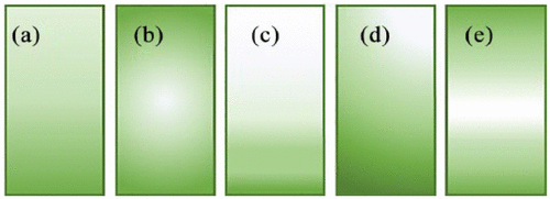
Classification of gradients according to their arrangements: (a) linear; (b) radial; (c) exponential; (d) orthogonal; (e) sigmoidal gradients, as representative of non-linear shape. Figure adapted from Smith Callahan
A gradient range of values over a given quality allows for a better defined more specific property at any given point. The researchers go on to explain how important gradients are in copying the anisotropy of tissue, along with acting as the basis of so many ‘phenomena’ behind cellular and bacterial results. They explore semi-immersion, diffusion, compositional topography, selective irradiation, and microfluidic devices.
“… the gradient is usually continuous, with smooth variation of the chemical or physical properties within the system. Both methods are suitable for the fabrication of scaffolds for the regeneration of a single tissue (bone, cartilage, skin, tendons, nerves, etc.) or for interface tissue engineering (cartilage-to-bone and tendon-to-bone, among others),” state the scientists.
Rapid prototyping, via 3D printing, allows for complex geometries to be created and easily modified. For studying bioprinting, the researchers used scans of target bone structure from a patient who had a scan performed. The scan was converted to 3D data, and a radial porosity gradient was formed.
“Biochemical gradients are more complex to achieve with bioprinting. Indeed, changes in the chemical composition of printed scaffolds imply the need to use different bioinks during the printing process,” stated the researchers.
Four bioinks were created and then printed to replicate both the extracellular matrix and vessel structures:
- Polydimethylsiloxane
- Methacryloyl-gelatin
- Pluronic F127 (a triblock poloxamer)
- Methacryloyl-gelatin loaded with cells
Scaffolds originating from gelatin and fibrinogen showed much better promise, but the researchers noted that printing times were ‘severely prolonged by the numerous switch times.’ They noted that inks would require crosslinking after printing, meaning that the choice of biocompatible materials is seriously restricted.
“Until now, the main application of graded structures is confined to studying the effect of each varying parameter over cell activities. Moving to bioinspired structures for regenerative medicine, some examples have been reported for graded scaffolds in bone and cartilage regeneration. However, the potential of engineering graded materials is underestimated in many applicative fields, such as in-vitro models and soft-to-hard regenerative medicine,” concluded the researchers.
“The characterization techniques should be expanded to meet the need to examine the peculiarity of graded materials and validate the productive methods. An increasing knowledge of the production and characterization of graded structures will allow new scenarios for engineering bioinspired materials to be explored.”
Find out more about other stories we have followed about scaffoldings and bioprinting, to include the creation of 3D printed devices. What do you think of this news? Let us know your thoughts! Join the discussion of this and other 3D printing topics at 3DPrintBoard.com.
[Source / Images: Engineering Biological Gradients]
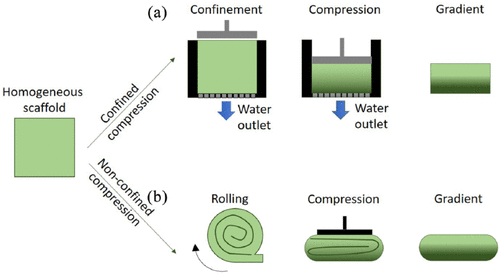
Confined (a) or non-confined (b) plastic compression can be exploited to induce gradients in the porosity and mechanical properties of elastic polymers. The homogeneous scaffold is compressed, in a molder or after rolling, with constant or variable load to induce alterations in the polymeric network and a macroscopic plastic deformation.