New Facility for Bioengineering Research Opens in Los Angeles

In a world eager to solve the problem of rejection in organ transplantation, a young American scientist developed a breakthrough test in 1964 that would help establish the compatibility of tissue types between organ donors and patients in need of transplants. Even though, today, efforts to meet organ transplant demand are shifting toward the field of bioengineering, as researchers search for ways to recreate complex organs with patient-derived cells, the legacy of that scientist, Paul Ichiro Terasaki, continues to inspire discoveries in transplant medicine through his philanthropic ventures.

The Terasaki Institute for Biomedical Innovation (TIBI), a nonprofit research organization established by Terasaki, professor emeritus of surgery at the David Geffen School of Medicine at the University of California Los Angeles (UCLA), will open the doors to a new facility in 2022. The newly-acquired addition will house interdisciplinary research in bioengineering, micro- and nanoscale technologies to enable transformative biomedical innovation as part of continuing research to solve the biggest problems related to organ transplantation and beyond.

Earlier this month, the Terasaki Institute announced the revamping of a building in the Woodland Hills area of the city of Los Angeles. Once home to the Weider Health and Fitness Center, created by bodybuilder and entrepreneur Joe Weider, the two-story building will be custom-designed to house the latest technology in cutting-edge research and will provide 50,000 square feet of floor space for up to 200 employees.

Located just 22 miles north of the original Terasaki Institute facilities in Westwood, the new space devoted to laboratory research will be designed to accommodate multiple teams of scientists, who will be developing bioengineered systems, devices, and other products with several biomedical applications. This new facility will be fully equipped to enable such technologies as tissue engineering and regeneration, biofabrication using 3D printing, nano- and micro-engineering, stem cell engineering, and the creation of human organs on chips.

When the new facility is inaugurated, with the renovation of the building set to begin in fall 2020, it will become the Terasaki Institute’s third research facility. In addition to the ample space and unique design features of the laboratory, the new facility will include in-house technology translation capabilities to be able to build prototypes and scale models of devices engineered by the institute. It will also be able to accommodate meetings, seminars, and conferences to further the education and exchange of ideas among its researchers and collaborators.

“I’m very excited about the addition of the new building to the Terasaki Institute. I believe that this addition will give us needed research space to bring together a number of leading scientists in our efforts to develop the next generation of biomedical innovations,” said Terasaki Institute’s new director and CEO, Ali Khademhosseini. “I’m particularly excited about furthering the great legacy of the Weider family and the building’s history in promoting health and fitness by focusing on individualized cures and diagnostics.”

Previously at Harvard Medical School, the Wyss Institute for Biologically Inspired Engineering, and most recently at UCLA Bioengineering, Khademhosseini has been an influential figure in pushing bioengineering forward. His research in regenerative medicine, tissue engineering, and micro- and nanotechnologies for the treatment of diseases has been related to advancements that allow reprogramming of adult cells to become progenitors, as well as editing genes. The bioengineer has also created a technique that uses a specially adapted 3D printer that could help advance the field of regenerative medicine by making it possible to 3D print complex artificial tissues on demand. He has also established the Khademhosseini Lab, an industry-leading tissue engineering lab that is co-sponsored by both MIT and Harvard and acts as a strategic partner to 3D bioprinting startup BioBots.

Ali Khademhosseini (Image: Ali Khademhosseini)

Stewart Han, president of the Terasaki Institute, has been working hard overseeing the planning and renovation of the new building: “It is exciting to be able to create a brand-new laboratory and research facility from the ground up, and it will greatly enhance our research capabilities when it’s completed. We also know that the new building will facilitate the future growth of our institute.

Founded in 2001, the Terasaki Institute was made possible through an endowment from the late Paul Terasaki, and it is expected to continue leveraging scientific advancements that enable an understanding of personalized medicine, from the macroscale of human tissues down to the microscale of genes, as well as to create technological solutions for some of the most pressing medical problems of our time.

Paul Terasaki in front of the Terasaki Life Sciences Building UCLA. (Image: Leslie Barton/UCLA)

“The board of the Terasaki Institute is very excited about the purchase of the new building in Woodland Hills, and we look forward to developing it into a world-class biomedical research center,” said board chair and diagnostic radiology specialist Keith Terasaki. “My father, the late Paul I. Terasaki, started the Terasaki Institute in hopes that it will make impactful discoveries in medical research. This new research facility will enable us to do so.”

To the field of transplant surgery, transplant pioneer Paul Terasaki enabled a broad understanding of organ transplant outcomes around the world. More than 70 years after his original discovery, patients still rely on organ donor transplants and the fundamentals of Terasaki’s laboratory developed tissue typing tests are still used today for the determination of transplant compatibility. Nonetheless, the Terasaki Institute envisions a world where personalized medicine is available to all. So, as the researchers at the institute continue to address the challenges that can finally advance the field of organ transplants from human donors to bioengineered artificial organs, they might bridge the gap between sickness and health. With one of the most productive 3D printing researchers as director, Khademhosseini, and a new facility to further explore biofabrication technology, we can expect to hear much more from the Terasaki Institute in years to come.

The post New Facility for Bioengineering Research Opens in Los Angeles appeared first on 3DPrint.com | The Voice of 3D Printing / Additive Manufacturing.

A Guide to Bioprinting: Understanding a Booming Industry

The success of bioprinting could become the key enabler that personalized medicine, tissue engineering, and regenerative medicine need to become a part of medical arsenals. Breakthroughs in bioprinting will enable faster and more efficient patient care and recovery. Biofabrication could be used to reshape the foundations of drug development, medicine, cosmetics, organ transplantation, and many other fields. It will transform the way doctors repair damaged ligaments, recreate tissues, and even reproduce the layers of the skin.

We are entering an era of bioprinting revolution. But to understand the role that bioprinting will play in the future, it is important to look back at how early discoveries in the field provided a strong basis to push its capability forward:

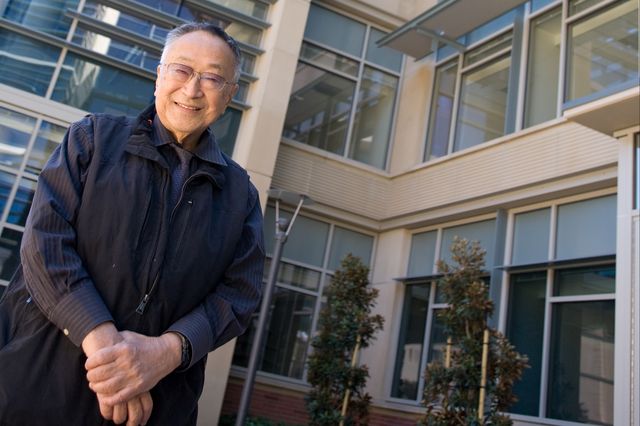
  • Back in the ’90s, Anthony Atala, pediatric surgeon, urologist, and director of the Wake Forest Institute for Regenerative Medicine (WFIRM) in North Carolina, created by hand bladders, skin, cartilage, urethra, muscle, and vaginal organs. By the end of that decade, the Institute used a 3D printer to build a synthetic scaffold of a human bladder, which they then coated with cells taken from their patients and implanted it, preparing the stage for bioprinting.
  • Then in 2002, scientists from Harvard University printed two-inch-long mini-kidneys capable of filtering blood that was then transplanted back into genetically identical cows, where they started making urine. The novel research raised the prospect of using stem cells taken from human patients with kidney failure to create new organs for transplant.
  • However, it wasn’t until 2003, when bioengineering professor Thomas Boland adapted a Hewlett-Packard inkjet printer in his lab at Clemson University to begin printing a bioink made of living bovine cells suspended in the cell-culture medium, that bioprinting began to materialize. This led to the creation of the world’s first 3D bioprinter, capable of creating living tissue from a solution of cells, nutrients, and other bio-compatible substances.
picture os a man using the bioprinter on a limb

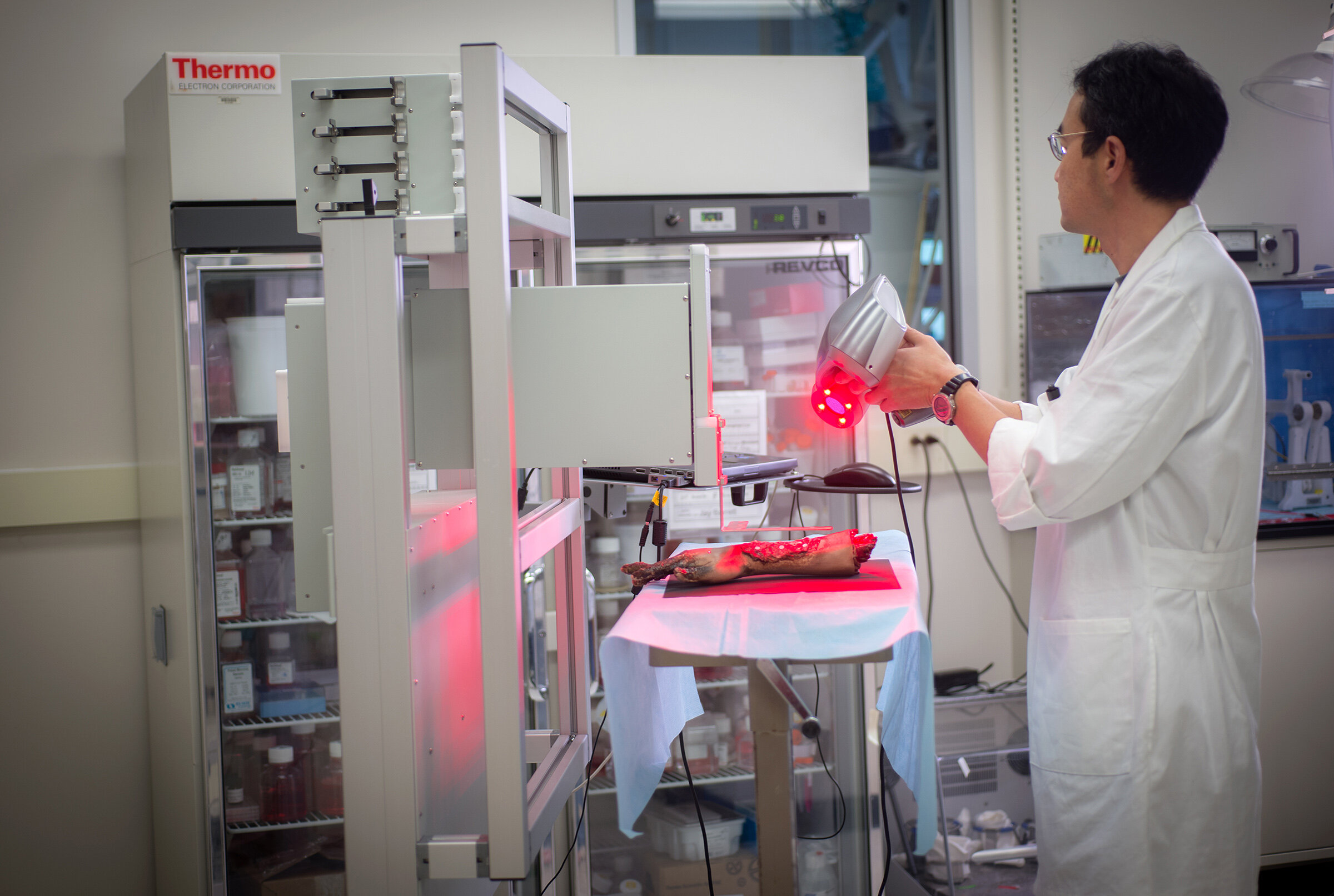
A member of WFIRM team operates with the bioprinter (Image: WFIRM)

Twenty years later, researchers still face challenges as they continue working with bioprinters and bioinks. Even though there has been an increasing adoption of the technology, the extent of its potential has not been fully exploited. From choosing the bioinks to actually bioprinting human tissues and organs, this new field is quickly becoming the go-to technology that bioengineers, researchers, and hospitals need to evolve from lengthy and cumbersome manual work to scalable and replicable results.

How Well Do you Know Bioprinters? (or How Bioprinters Work and Who Makes Them)

Bioprinters work by extruding cells and other biomaterials contained in bioinks, from syringes that deposit the material layer by layer to create different types of tissues or organ-like constructs. The technology behind the bioprinters vary. Nonetheless, to date, the three main and most popular bioprinting technologies are extrusion, inkjet, and laser-based bioprinting. Some mainstream examples are:

  • Some manufacturers, like Cellink or Allevi, use pneumatic-driven extrusion systems that pump high-pressure air in a cartridge to force bioinks to flow through a nozzle. 
  • Other fabrication systems, such as the one designed by Poietis has laser-assisted bioprinting that allows cells to be positioned in three dimensions with micrometric resolution and precision to design living tissue.
  • Another type of bioprinting technology uses a stereolithography-based bioprinting platform. Vendors using this process include Volumetric and Cellink’s jointly produced Lumen X projection stereolithography based bioprinter.
  • Another project that could revolutionize the way surgical procedures are performed is handheld bioprinters; these systems enable surgeons to deploy cells — or material to aid in cellular growth — directly into a defect site in the body, such as severely burnt skin, corneal ulcerations or bone. One of the most talked-about handheld bioprinters has been Australia’s University of Wollongong BioPen, allow surgeons to repair damaged bone and cartilage by “drawing” new cells directly onto bone in the middle of a surgical procedure. Although still in pre-clinical trials, these devices have attracted the attention of healthcare practitioners due to its versatility.

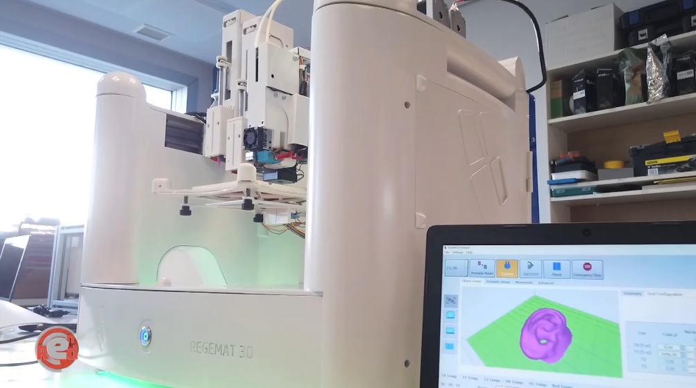
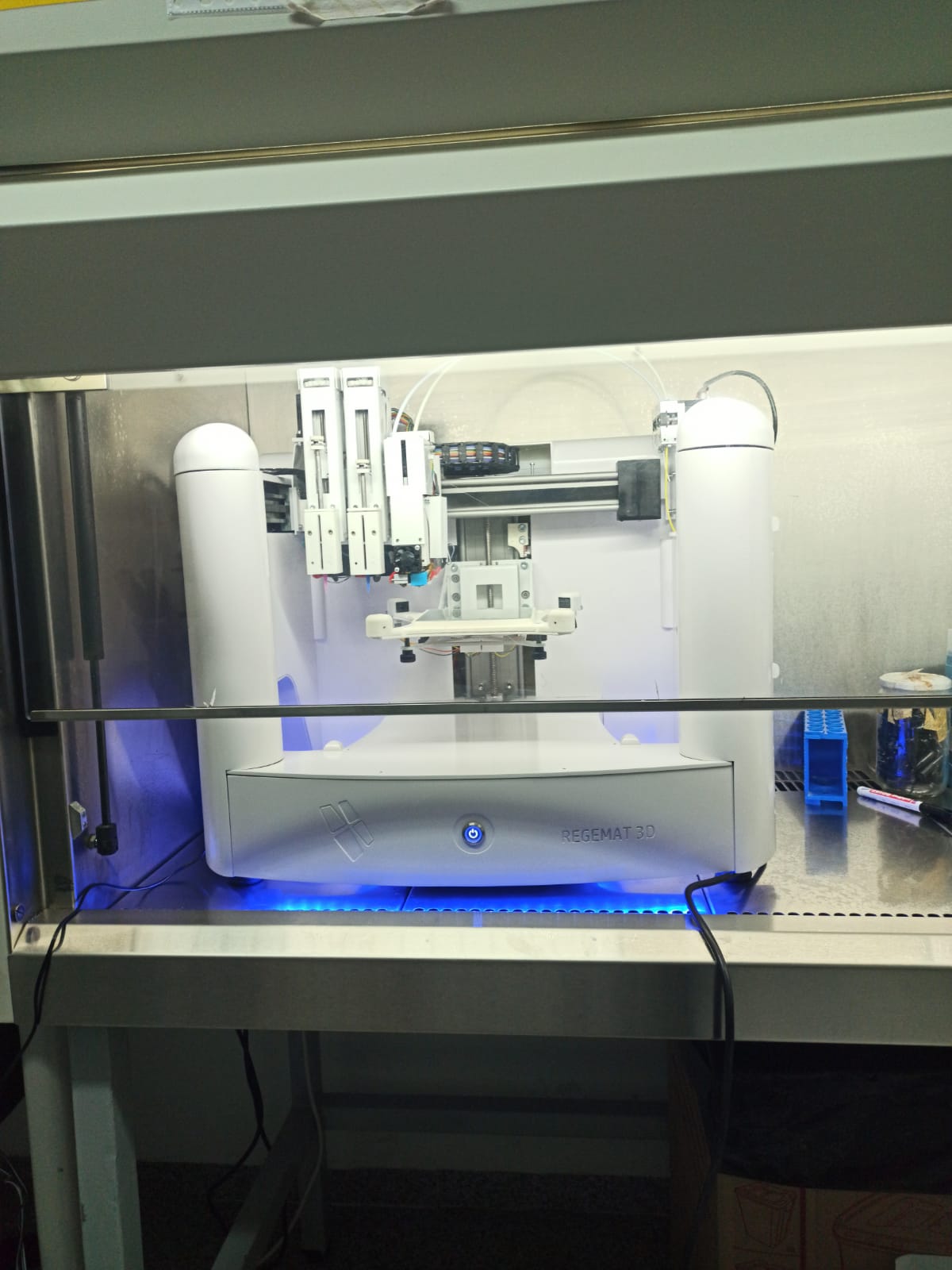
A few of the main manufacturers supplying the market include 3D Bioprinting Solutions, Allevi, Aspect Biosystems, Cellink, nScryptregenHuInventiaRegemat3DPoietis, and more. Last year, 3DPrint.com counted 111 established bioprinting firms around the world. Mapping the companies that make up this industry is a good starting point to understand the bioprinting ecosystem, determine where most companies have established their headquarters and learn more about potential hubs, like the one in San Francisco.

Types of Bioinks

3D bioprinters use bioinks. Bioinks are substances made of living cells that can be used for 3D printing complex tissue models — they mimic an extracellular matrix environment to support the adhesion, proliferation, and differentiation of living cells. Choosing which bioink to use can be challenging. To date, we have witnessed researchers using bioinks based on several biomaterials, such as alginate, gelatin, collagen, silk, hyaluronic acid, even some synthetic-biomaterials-based-bioinks.

The promise of hydrogels. A macromolecular polymer gel constructed of a network of cross-linked polymer chains, hydrogels are able to meet the stringent requirements of cells and are the basis of almost all bioink formulations. As stated in “Engineering Hydrogels for Biofabrication”, published in Advanced Materials, “hydrogels are particularly attractive for biofabrication as they recapitulate several features of the natural extracellular matrix and allow cell encapsulation in a highly hydrated mechanically supportive threedimensional environment.” This makes hydrogel-based bioinks a very promising choice for many researchers and bioengineers.

Bioinks from patients’ cells. The biomaterials can also use a patient’s own cells, adult stem cells, manipulating them to recreate the required tissue. The source of the cells varies depending on what researchers are bioprinting. For example, in the case of Alzheimer’s disease, experts at the University of Victoria in Canada, have bioprinted neural tissues using stem cells as a tool for screening drug targets for the disease. The ability to program patient-specific cells is the beginning of customized bioprinting since the unlimited potential of these cells can be used to regenerate or repair damaged tissue. 

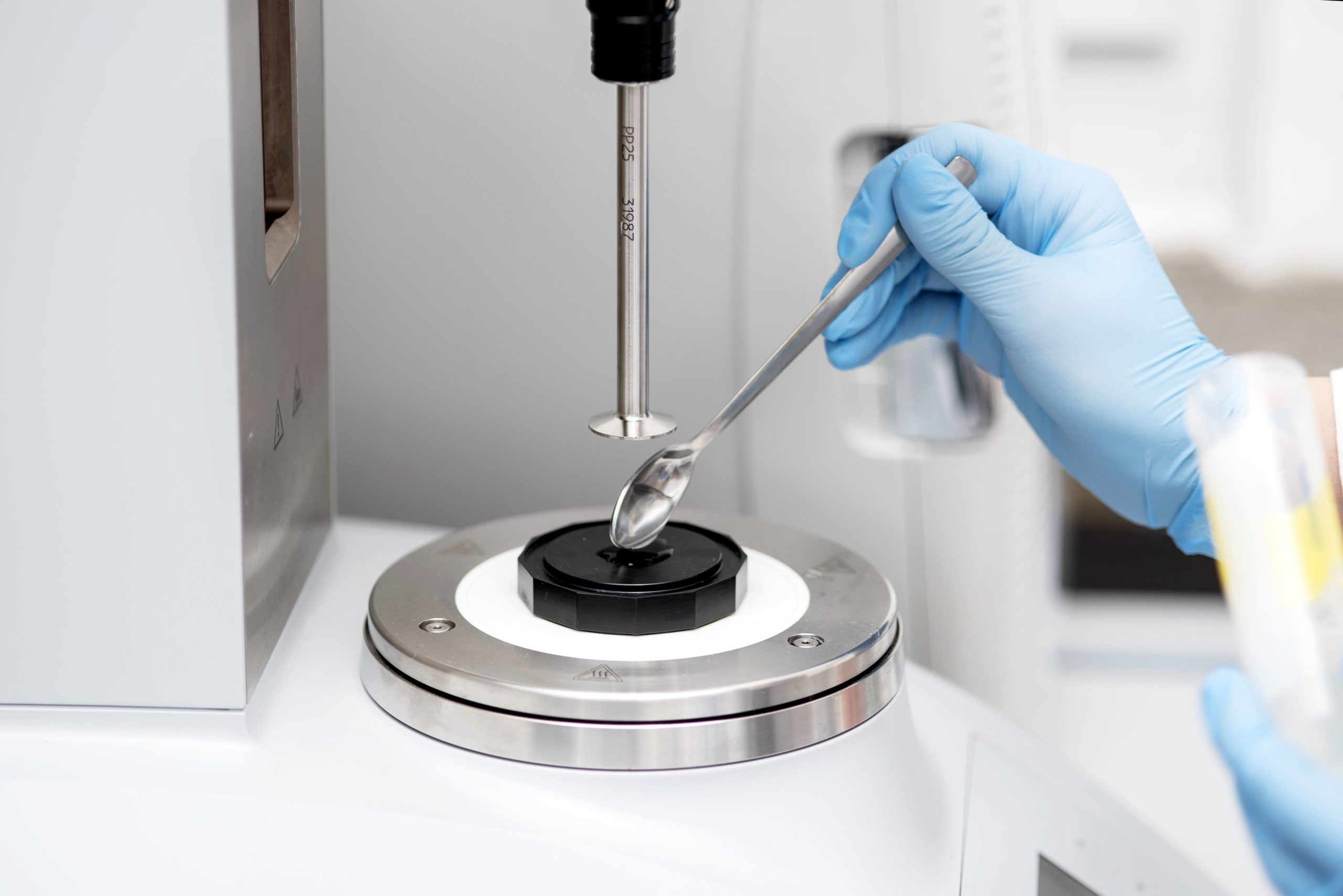
Polbionica’s bioinks (Image: Polbionica)

What is Bioprinting Good For?

What is most exciting about bioprinting, are the many ways that doctors and researchers are using currently available devices in the market or are creating their own systems to facilitate new processes and applications. The orchestrated interaction between machine and user has led to innovation that could reinvent the world of tissue engineering.

Moving oncology forward:  Bioprinting is being employed in the battle against cancer, whereby scientists create tumor models for research. Modeling cancer using 2D cell cultures fails to accurately replicate the microenvironment of tumors. This is why scientists have turned to biofabrication tools to make three-dimensional models that mimic the intricate in vivo tumors. These models help test anticancer drugs; aid scientists in understanding the underlying causes of metastasis, and can even personalize treatment for individual cancer patients. There have been plenty of initiatives that apply bioprinting to oncology. These range from immersion bioprinting of human organoids to printing cancer tissues in 3D

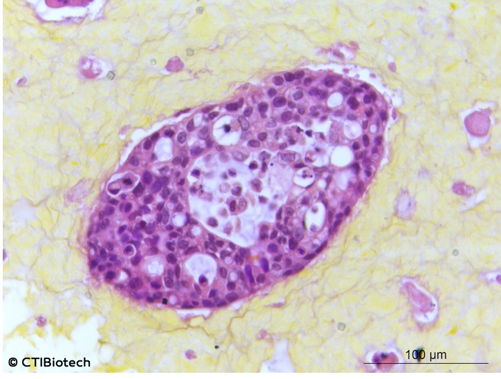
Microtumors (Image: CTI Biotech)

The market for oncology-oriented bioprinting seems sure to grow. The number of patients suffering from the disease continues to go up. In 2018 alone there were 17 million new cases of cancer worldwide, and projections suggest that there will be 27.5 million new cases of cancer each year by 2040. What that effectively means is that we are witnessing an increase in oncology bioengineering research and whether it is for glioblastoma, bone cancer tumors, or lung models with tumors, the implications can be profound since the ability to use bioinks and bioprinters to create tumors frees researchers of the many ethical concerns associated with testing as well as reduces the costs associated with such research activities. 

Building scaffolds: Probably the most important practical use for bioprinting at the present time is in regenerative medicine. For instance, in 2019, researchers from North Carolina State University (NCSU) and the University of North Carolina at Chapel Hill created a 3D biomedical fiber printer used to create biocompatible scaffolds. Also, Harvard researchers working in Jennifer Lewis’ Lab at Harvard´s Wyss Institute for Biologically Inspired Engineering and John A. Paulson School of Engineering and Applied Sciences (SEAS), came up with a much talked about breakthrough new technique, the SWIFT method, that allows 3D printing to focus on creating the vessels necessary to support a living tissue construct. A team of researchers at Texas A&M University have even developed a 3D printable hydrogel bioink containing mineral nanoparticles that can deliver protein therapeutics to control cell behavior.

Limitations of Bioprinting

Though we heard it many times before, knowing that someday 3D printed artificial organs could eliminate the need for an organ donor waiting list is comforting. Creating personalized replacement organs sounds like the solve-all solution to the organ shortage crisis, yet, a functional organ compatible for human implantation may be decades away. Today, 3D printed organs are still raw to be used for transplantation and lack the vasculature required to function within the human body.

Creation. Last year, mainstream news outlets headlined a story about researchers who had 3D printed a heart. However, the published scientific paper behind that story described how a group of scientists from Tel Aviv, Israel, created bioink out of heart cells and other materials from a patient, and were able to develop cardiac patches and ultimately, 3D print comprehensive tissue structures that include whole hearts. The tissue was shaped in the form of a tiny heart that was kept alive in a nutrient solution. The paper expresses how this development could not function like a real heart since the cells in the construct can contract, but don’t yet have the ability to pump.

This was certainly not the first heart to be 3D printed, yet Tal Dvir, who led the project at Tel Aviv University’s School of Molecular Cell Biology and Biotechnology, indicated that never before had it resulted in an organ “with cells or with blood vessels.” It was an amazing breakthrough for the field, and it proves that biotechnology has made significant advances, but it is still a long way from creating organs that can be transplanted to people, considering that the vasculature — the network of blood vessels that feeds the organ — remains a challenge, but scientists are determined to troubleshoot these issues.

So, no matter how enticing the idea of successfully bioprinted organs sound, stories like this remind us to keep the hype in check, making the work of news outlets fundamental for reporting advancements in research and medical breakthroughs (which usually take much more time).

Where to Next?

Organ bioprinting. The application of 3D bioprinting will be a game-changer in medicine, as the machines successfully replicate tissues and organs, build muscles and cartilage, and enable the adoption of customized medicine. The long-term dream for bioprinting has always been the routine printing of body organs. Current ongoing projects include Michal Wszola’s 3D bioprinted bionic pancreas or Organovo’s 3D bioprinted liver.

The space frontier. Bioprinting in space could hold the key to developing fully functional organs. This is because bioprinting without gravity allows organs to grow without the need for scaffolds. The National Aeronautics and Space Administration (NASA) considers that terrestrial gravitation represents a significant limitation, while a gravity-free environment, magnetic and diamagnetic levitation will allow for biofabrication of 3D tissue constructs with a scaffold-free and even nozzle-free approach. Some bioprinters have already been launched and used in space.  This includes nScrypt and Techshot’s BioFabrication Facility (BFF), or the Organaut 3D bioprinter created by Russian biotech firm 3D Bioprinting Solutions and Roscosmos, the Russian state corporation responsible for space flights.

Healthcare at its best. More prosaically, biomaterials specialist and a professor of biofabrication at Queensland University of Technology, Australia, Mia Woodruff, has been advocating the hospitals of the future for years. She has an exciting vision of a future where the fabrication of patient-specific replacement tissue and organs is safe, cost-effective, and routine. Though perhaps years from happening, her vision is in tune with what many think bioprinting could become, that is, with enough researchers, companies, and funding.

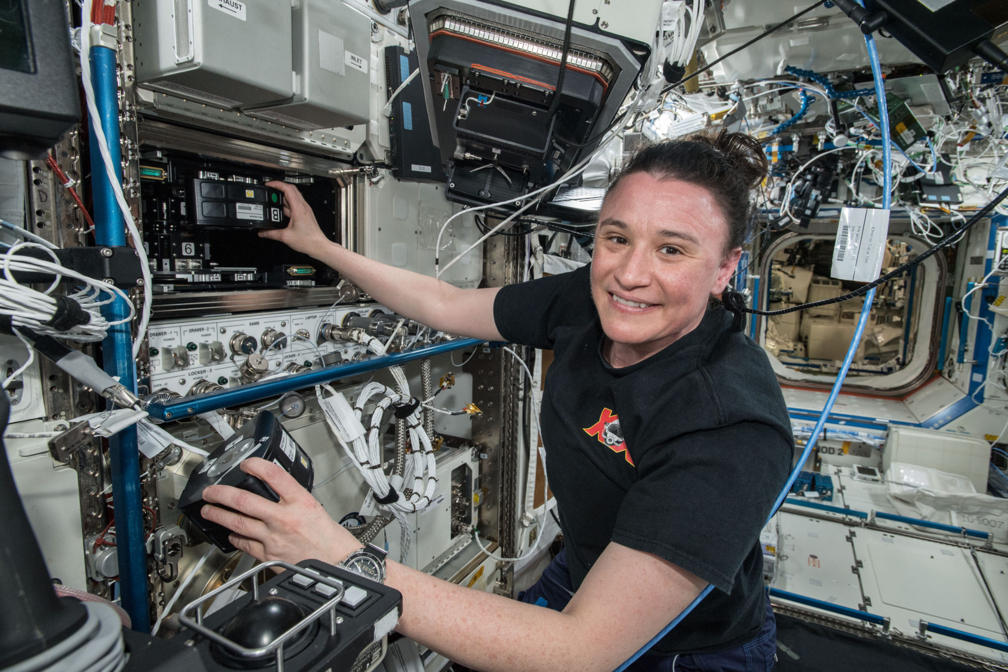
An astronaut aboard the ISS using Techshot’s BioFabrication Facility or BFF (Image: Techshot/NASA)

Coming regulation. Back here on Earth, there will be a growing need for common guidelines for bioprinting to make the process more standardized. In the EU, for example, there currently no particular regulatory regime governing the whole bioprinting process, but piecemeal legislation is relevant in relation to tissue engineering and regenerative medicine. While the Food and Drug Administration (FDA) of the United States plans to review the regulatory issues related to the bioprinting of biological, cellular and tissue-based products in order to determine whether additional guidance is needed beyond the recently released regulatory framework on regenerative medicine medical products.

As the development of the technology strongly advances and proves successful for researchers, we will surely continue to observe brilliant minds perfecting devices and biomaterials, envisioning new systems for future needs, especially as startups emerge out of universities and research institutes, and established companies upgrade their machines to face the limitations we previously addressed in this article.

Still, there is a long way to go, what was largely built so far is a very promising technology. For instance, fully functional organ fabrication for transplantation might take decades. Nonetheless, the unquestionable contribution of bioprinting to so many fields remains an incentive to invest in this area to overcome medical challenges and to move the healthcare industry in a different path, where technology will not only aid in curing diseases but also guiding people by helping them stay healthy, recognizing symptoms early and personalizing solutions in real-time. 

The post A Guide to Bioprinting: Understanding a Booming Industry appeared first on 3DPrint.com | The Voice of 3D Printing / Additive Manufacturing.

Cell Culture Bioreactor for Tissue Engineering

Researchers from the US and Portugal are refining tissue engineering applications further, releasing the findings of their study in the recently published ‘A Multimodal Stimulation Cell Culture Bioreactor for Tissue Engineering: A Numerical Modelling Approach.’ In creating a new bioreactor for 3D printing, the authors worked to promote reproducibility and optimization, fabricating a design not possible using conventional techniques.

In most bioprinting, cells are seeded onto scaffolds—also the source of much study, whether in regard to new techniques, enhancements, or interface engineering—and then researchers must hope for viability. Promoting cell growth and sustainability is one of the greatest challenges in any type of bioprinting, and often devices like perfusion flow bioreactors are used in replacing culture mediums to remove toxins. In some cases, they are also used for mechanical stimulation through fluid flow shear stress (FFSS). Electric field stimulation devices can also be easily used to encourage cells to grow, mature, and differentiate.

The bioreactor used here was part of a previous study conducted by the authors, but now updated (using SOLIDWORKS 2018 Student Edition ) to include capabilities in E-Field stimulation and fluid flow mechanical stimulation. 3D design was performed in SOLIDWORKS 2018 Student Edition. Assessment of the new design was part of this study, leaving the team to create a numerical finite element analysis (FEA) of the model.

With FEA, the researchers could project input conditions for the bioreactor, further enhancing effectiveness of cell stimulation, determined from in vitro data. Overall, in vitro tests can offer ‘essential’ information for confirming ranges of multimodal stimulation—projected via numerical studies.

Numerical finite element analysis (FEA) analysis of the proposed bioreactor design with a DC electric stimulation parallel plate capacitor set-up with lateral and top slice views. The three top views represent the ROI upper slice (T1), the ROI middle plane slice (T2) and the ROI bottom slice (T3). (a) Electric potential distribution predicted in the bioreactor due to DC stimulation. (b) E-Field magnitude distribution predicted for the same electric DC stimulation conditions.

Numerical FEA analysis of the proposed bioreactor design for a laminar perfusion flow with lateral and top slice views. The three top views represent the ROI upper slice (T1), the ROI middle plane slice (T2) and the ROI bottom slice (T3). (a) Pressure distribution predicted considering applied inlets velocity of 0.003 m/s and a outlet pressure of 0 Pa. (b) Fluid velocity distribution predicted for the same inlet/outlet conditions. The velocity distribution at the ROI middle plane slice is presented in more detail in a top view inset at the right of the slice plane.

“Electrical and mechanical stimulation conditions in the region-of-interest (ROI) were considered for bone cell stimulation optimization, according with reference values obtained from two previous in vitro studies on bone cell stimulation, one applying mechanical stimulation, and the other using E-Field stimulation,” explained the authors.

Novel bioreactor design: (a) Vertical cut view of the bioreactor design, where the parallel electrodes set up, the upper and bottom inlets and the inlet flow splitters can be observed. (b) Horizontal cut view of the bioreactor design, where the radial outlet system can be observed. The green regions represent the region-of-interest (ROI) where the scaffold will be placed, represented by a cylinder with 4 mm of height and a diameter of 10 mm. (c) CAD bioreactor design assembled in frontal view, the main outlet hole is visible in the middle. (d) CAD bioreactor design assembled in lateral view, showing both electrode connector wires (in brown).

Flow splitters were added between the inlet and the scaffold, resulting in ‘indirect flow prevalence.’ Inlets and outlets were fitted with hose joiners, connecting them to the perfusion pump. The cell culture chamber was separated into two different areas for cell cultures and cell seeding exercises. Materials for use in the platform were required to be non-toxic and suitable according to ISO 10993-5 standards.

Bioreactor geometry volume mesh created using COMSOL Multiphysics, with 1.9 × 10⁶ elements, and an average element quality of 0.65.

“Accordingly, in the direct contact test, cells cultured in contact with all the materials presented normal fibroblast morphology with no evidence of any inhibition halo effect or cell death. According to the cytotoxicity tests results, all candidate materials are suitable for our bioreactor AM fabrication,” concluded the researchers. “We will consider C8 and PETG as materials of interest for future design fabrication. C8 is a new material with good layer adhesion and surface quality, which are key features for the perfusion flow. The C8 supplier datasheet reveals that this material has a higher tensile strength than ABS, resulting in improved mechanical characteristics, which are important for the overall robustness of the bioreactor to withstand the tightness of pressure chambers.”

Cytotoxicity assay with L929 mouse fibroblast according to ISO 10993-5 standards: (a) indirect contact (MTT protocol); (b) direct contact (digital images of the material samples and the negative and positive controls, fresh culture medium and Latex, respectively). A one-way ANOVA with no corrections for multiple comparisons (Fisher’s test) statistical analysis was performed using GraphPad Prism6.

“A design–numerical modelling approach will be essential to understand the underlying biophysical effects of electric and mechanical stimuli in cell cultures and can be a powerful tool for standardization of stimulation protocols considering different bioreactor designs and specific TE outcomes.”

What do you think of this news? Let us know your thoughts! Join the discussion of this and other 3D printing topics at 3DPrintBoard.com.

[Source / Images: ‘A Multimodal Stimulation Cell Culture Bioreactor for Tissue Engineering: A Numerical Modelling Approach’]

The post Cell Culture Bioreactor for Tissue Engineering appeared first on 3DPrint.com | The Voice of 3D Printing / Additive Manufacturing.

3D Printing: Successful Scaffolds in Bone Regeneration

In ‘Comprehensive Review on Full Bone Regeneration through 3D Printing Approaches,’ the authors review new developments and solutions in tissue engineering for the formation of cells, as well as proposing an optimized temporary support geometry for treatment.

Fused Filament Fabrication (FFF) process.

Bone regeneration continues to challenge researchers in their work as well as medical professionals attempting to improve patient treatment:

“Many research groups have been working on bone regeneration for over 10 years, but this has not led to effective therapy in a clinical setting. If it was successful, it would enhance the quality of life for millions of people and significantly reduce the absence to work due to fractures which are considered the second higher cause of working day lost,” state the authors.

“When there are fractures with a bone defect exceeding a critical size, the bone is not able to self-regenerate and, therefore, requires the use of a temporary implant (natural and/or synthetic) to serve as support and cells to help bone regeneration. In this way, tissue engineering (TE) has emerged.”

While scaffolds are used in tissue engineering for transporting nutrients and secretion of waste, the cells must be able to imitate true tissue biology, morphology, and functionality.

Exploring the usefulness of temporary implants, the authors state that in tissue engineering for patients, it is first critical to examine native bone tissue and mechanical properties.

Human long bone properties.

3D printed implants must be able to sustain cell viability in a secure environment, and scaffolds must possess suitable elasticity for matching regular bone. High porosity is desired in most tissue engineering, along with the use of materials that are not only biocompatible but also biologically active. During trials, animal models of fractures are often used in vivo before procedures are attempted on humans.

“Animal studies are needed to understand bone regeneration. Variables such as the amount of bone formation and its kinetics, mechanical properties and safety obtained by the scaffold, including the presence of toxic degradation in different organs and in terms of inflammatory response need to be understood in detail,” explained the researchers.

“However, bone fractures performed in animals do not represent the complexity of healing human fractures. The potential of each different type of cells both in vitro and in vivo plays here a key role.”

Even more interesting though, the authors point out that growth factors are unnecessary, with cells showing the potential to secrete optimal extracellular matrix (ECM) components.

“In vitro studies are advantageous because they offer a controlled environment to experimental test molecular and cellular hypotheses,” stated the researchers. “However, cells cultured in vitro are not replicates of their in vivo counterparts.”

While tissue engineering can be a delicate process overall in terms of working to keep cells alive, bone generation is particularly challenging—and scaffolds must be relied on to maintain the same role as tissue. Biomaterials must be able to mimic the natural environment, along with possessing identical mechanical properties of the initial bone. Appropriate levels of degradation are critical for bone regeneration, and are also dependent on corrosion resistance and materials.

Characteristics of the different materials used to produce a scaffold.

Suitable materials include poly(ε-caprolactone) (PCL) or polylactic acid (PLA), both approved by the FDA and offering stability, biocompatibility, and biodegradability. Scaffolds must be osteoinductive for sustaining cells as well as being osteoconductive, providing growth. They must also serve to:

  • Fill bone defect
  • Ensure pore connectivity
  • Encourage bone formation
  • Promote bone growth

Natural organization of long bones.

Designed in SolidWorks, the structures exhibited ‘superior advantages’ over what could be produced conventionally.

“Considering all types of materials available, associated with the desired bone regeneration and the use of synthetic polymers, as PCL or PLA, combined with collagen type I for the trabecular region and Hap for cortical region, seems to be the best strategy to follow,” concluded the researchers.

“Among the most commonly used bioreactors for bone regeneration, perfusion bioreactors appear as the most suitable, because it improves osteogenic proliferation and differentiation due to improved mass transfer and adequate shear stress. When making a design proposal for bone regeneration, it is necessary to study the mechanical effects, such as stress and tension, and link them.”

Cylindrical scaffold

DNA chain-inspired cylindrical scaffold.

Tissue engineering continues to be an enormous area of study, from seeding human dermal fibroblasts, promoting hydrogel microenvironments, to bioprinting structures for soft tissue engineering applications.

Scaffold requirements in terms of response (left) and what should be taken into account (right) (adapted from [106]).

What do you think of this news? Let us know your thoughts! Join the discussion of this and other 3D printing topics at 3DPrintBoard.com.
[Source / Images: ‘Comprehensive Review on Full Bone Regeneration through 3D Printing Approaches’]

The post 3D Printing: Successful Scaffolds in Bone Regeneration appeared first on 3DPrint.com | The Voice of 3D Printing / Additive Manufacturing.

Denmark: 3D Printing Conductive Hydrogels for Medical Applications

In the recently published ‘Electrically Conducting Hydrogels for Health care: Concept, Fabrication Methods, and Applications,’ Shweta Agarwala of the Department of Engineering at Aarhus University in Denmark researchers 3D printing techniques in the medical realm, offering a review of conductive hydrogels.

As 3D printing began to infiltrate the mainstream, industries such as automotive, aerospace, and construction have been positively impacted—and now, medical applications have gone beyond 3D models and devices as scientists continue to make huge strides in bioprinting. Scaffolds are a common structure used in tissue engineering featuring a variety of different hydrogels; however, they can also be used for a wide range of applications today, from smart wearables to biosensors, implants, and avenues to wound management.

Hydrogels are attractive for use in research and other applications due to:

  • Ideal extracellular matrix (ECM)
  • Cell support
  • Biocompatibility
  • Natural and synthetic hydrophilic polymer chains offering high absorption of water

“Although hydrogels have found niche application in tissue engineering, they are inherently insulating by nature. Recent research has shown that hydrogels not only possess necessary characteristics to support biological species but can also interface with electrical circuitry if modified,” states Agarwala. “Hence, research on conducting hydrogels have gained widespread interest for applications such as health recording electrodes.”

Schematic illustration of conducting hydrogels, their components and applications.

Agarwala notes that in general, the conductivity associated with hydrogels is ionic conductivity.

Summary of the material composites and their electrical conductivities achieved to make conducting hydrogels.

“The contribution from additives materials to overall conductivity in such cases is small. However, recent research efforts in this direction have shown promise in inducing electrical conductivity from the additive materials,” states Agarwala.

While the method most often used for aqueous compatible conducting materials is to use ultrasonic energy or heating, five other approaches are available:

  1. Hydrogel monomers with cross-linkers and nanoparticles are gelated together.
  2. Nanoparticles are physically embedded into the hydrogel matrix after gelation.
  3. Nanoparticle precursors are loaded into the gel.
  4. Cross-linking using nanoparticles forms hydrogels.
  5. Hydrogels are formed using nanoparticles, polymers, and other molecules.

Schematic diagram depicting various approaches to synthesize conducting hydrogel: (A) hydrogel monomers with cross-linkers and nanoparticles gelated together; (B) physically embedding nanoparticles into hydrogel matrix after gelation; (C) reactive nanoparticle formation aided by the hydrogel network where nanoparticle precursors are loaded in the gel; (D) cross-linking using nanoparticles to form hydrogel; and (E) hydrogel formation using nanoparticles, polymers, and other molecules.

One of the greatest benefits in 3D printing is that users are able to create much more complex geometries, along with enjoying enormous latitude in both design and customization, as well as being able to make projects faster, and make changes to them on demand. All of these benefits apply to why there has been such an increase in 3D printing conducting hydrogels.

Techniques usually rely on shear thinning, causing them to flow as pressure is applied, using a piezoelectric head.

“A piezoelectric material deforms on applying voltage or current. Thus, the orifice opening can be controlled by varying the voltage applied to the printer head. Inkjet printing creates small droplets (sub-micron volume), which are deposited on the surface,” states Agarwala. “Small volume of material deposition, as against large material ejection through extrusion, helps to print high-resolution constructs and scaffolds.”

“Ink development is considered one of the most important aspects of 3D printing. Hydrogel inks need to have the right rheological properties to fulfill the physical and mechanical needs of the orienting process.”

Sketch of (A) 3D bioplotting system (Reproduced with permission [65]) (B) digital light projector (DLP) 3D printing system to 3D print conducting hydrogel scaffolds (Reproduced with permission [74]), and (C) stereolithography process (Reproduced with permission [75]).

Such hydrogels have the potential to be used in sensor technology, drug delivery systems, and tissue engineering. A variety of composites have been used too, from graphene-chitosan to silica nanoparticles, silica alumina, but the author points out that commercialization of such manufacturing is ‘still far away,’ due to the many challenges involved.

“These materials are unable to follow the original design models, as the printed construct does not retain the original shape. Achieving functional gradients and hierarchical properties have also been challenging and new design approaches are being developed to tackle them,” concludes Agarwala. “

“The area of conducting hydrogels is still full of unresolved technological challenges, and thus provides researchers with opportunity for development, as this field is growing fast beyond its early stage. Improvement in the conductivity of the hydrogels may be one research direction, while incorporating new functionalities such as biodegradability and mechanical strength can open new avenues for applications. Innovation is also required in fabrication methods to allow varied composition of hydrogels to be laid down in desired fashion.”

What do you think of this news? Let us know your thoughts! Join the discussion of this and other 3D printing topics at 3DPrintBoard.com.

[Source / Images: ‘Electrically Conducting Hydrogels for Health care: Concept, Fabrication Methods, and Applications’]

The post Denmark: 3D Printing Conductive Hydrogels for Medical Applications appeared first on 3DPrint.com | The Voice of 3D Printing / Additive Manufacturing.

Researchers in Asia create implantable blood vessels using 3D cell printing

Researchers in Korea and Hong Kong have used a modified 3D cell printing technique to fabricate a biomimetic blood vessel that was successfully implanted in a living rat.  According to the researchers, their approach to tissue-engineered biomimetic blood vessels outlined in their study provides a promising route for the construction of durable small-diameter vascular grafts. […]

3D Printed Haversian Bone: Biomimicking for Cell Regeneration

Chinese researchers continue to take on the challenge of bone regeneration in tissue engineering, sharing their findings in the recently released ‘3D printing of Haversian bone—mimicking scaffolds for multicellular delivery in bone regeneration.’

The fabrication of scaffolds is at the center of successful bone regeneration, but this is an area of medicine that is still known to be difficult—despite a wide range of research projects performed already, from using variations in nanotubes to structures with controlled antibiotic release, alternative materials and coatings.

As the authors point out, while bioprinting is an area of medical research where scientists have made huge strides, there are several reasons that obstacles remain:

  • Complexity of hierarchical structures
  • Mechanical property requirements
  • Diversity of bone resident cells

Most regeneration occurs in cancellous bone, however, so this is what researchers must focus on for success in treating patients:

“Cancellous bone is a meshwork consisting of plate-like or rod-like structures at about 200-mm thickness,” explained the authors. “By one estimate, 80 percent of bone remodeling processes occur in cancellous bone. However, bone regeneration not only needs to reconstruct bone structure but also involves repairing other tissues like blood vessels or nerves.”

Biomimetic structures are showing great potential as ‘high-performance bone tissue engineering biomaterials,’ however, leading the research team to focus on the use of 3D printed Haversian bioceramic structures which can imitate bone in a ‘simple but versatile design.’

3D printing Haversian bone–mimicking scaffolds integrated with Haversian canals, Volkmann canals, and cancellous bone structure for delivery of osteogenic and angiogenic cells. Osteogenic cells were seeded in cancellous bone structure of scaffolds, and angiogenic cells were seeded on Haversian canals. The Haversian bone–mimicking structure–based multicellular delivery system contributed to the formation of new bone and new blood vessels.

Due to superior osteoconductivity and osteoinductivity, materials composed of akermanite were used in fabrication of five samples with varying measurements in canals and cancellous bone serving as meshwork.

3D printing of Haversian bone–mimicking bioceramic scaffolds with cortical bone and cancellous bone structure. Cortical bone structure contained Haversian canals and Volkmann canals. (A to E) Optical microscope images exhibited different diameters (D) and numbers (N) of Haversian canal indicated by magenta arrows (A) N = 8, D = 0.8 mm; (B) N = 8, D = 1.2 mm; (C) N = 8, D = 1.6 mm; (D) N = 4, D = 1.6 mm; and (E) N = 2, D = 1.6 mm. Scale bars, 1 mm. (a to e) Micro–computed tomography (CT) images show Volkmann canals (blue arrows) connecting Haversian canals in the interior of scaffolds. Scale bars, 1 mm. (F to J) SEM images presented the microstructure of the scaffolds.

“Interconnected Haversian canals were isolated from the cancellous bone structure for noncontact cell coculture,” stated the researchers. “Furthermore, the periphery and bottom of the scaffolds were sealed so that the scaffolds could be used for holding the seeded cells.”

Several different techniques were necessary in creating the scaffolds, to include using electrospinning and twin-screw extrusion, modular tissue engineering, and 3D printing. The team had the most success with DLP 3D printing, designing a series of structures with Haversian canals, Volkmann canals, and cancellous bone.

Haversian bone–mimicking bioceramic scaffolds for the HBMSC-HUVEC coculture system performed better in cell proliferation and angiogenic differentiation than monoculture. (A to D) CLSM images of HBMSCs seeded on the cancellous bone structure (A) and HUVECs seeded on the Haversian canal with different diameters, (B) D = 1.6 mm, (C) D = 1.2 mm, and (D) D= 0.8 mm. Scale bars, 100 m. (E to H) SEM images of (E) HBMSCs seeded on the cancellous bone structure and HUVECs seeded on the Haversian canal with different diameters of (F) 1.6 mm, (G) 1.2 mm, and (H) 0.8 mm. (I and J) The proliferation activity of HBMSC, HUVEC, and cocultured HBMSC-HUVEC seeded on scaffolds with different (I) diameters and (J) numbers of Haversian canals after culturing for 1, 3, 7, and 14 days. n = 6 replicates. (K and L) The osteogenic (K) and angiogenic (L) gene expression of HBMSC, HUVEC, Co-HBMSC (HBMSCs in HBMSC-HUVEC coculture), and Co-HUVEC (HUVECs in HBMSC-HUVEC coculture) for 3 days. n = 3 replicates. *P < 0.05, **P < 0.01, ***P < 0.001, $ P < 0.05, $$P < 0.01.

Haversian bone–mimicking bioceramic scaffold–based rBMSC-rSC coculture system performed better in cell proliferation and neurogenic differentiation
than monoculture. (A to D) The CLSM images of rBMSC in the (A) rBMSC monoculture group and rBMSC-rSC coculture group with the ratio of rBMSC to rSC being (B) 3:7, (C) 5:5, and (D) 7:3 seeded on the cancellous bone of scaffolds. Scale bars, 50 m. (E to H) The CLSM images of rSCs in (E) the rSC monoculture group and rBMSC-rSC coculture group with the ratio of rBMSCs to rSCs being (F) 3:7, (G) 5:5, and (H) 7:3 seeded on the Haversian canal of scaffolds.

“Here, we further established the Haversian bone–mimicking scaffold–based rBMSC-rSC coculture system with rBMSCs grown in cancellous bone structure and rSCs grown on Haversian canals. Our results indicated that the rBMSC-rSC coculture system exhibited a better proliferation and a higher expression of NGF, BDNF, TrkA, and S100 as compared to rSC monoculture,” concluded the researchers. “It was found that BMSCs could promote SC proliferation and were traditionally used to facilitate the recovery of sensory system.”

“Considering the clinical applications of Haversian bone–mimicking scaffolds, there are still some issues to be studied. First, more bone-resident cells such as osteoblasts, osteoclasts, and macrophages should be further considered in the coculture system. The mechanism of multicellular synergistic effects is not fully understood. Further studies are needed to identify the individual effects of coculture cells on the formation of new bone, blood vessels, and nerves in the Haversian bone–mimicking scaffold–based coculture system.”

What do you think of this news? Let us know your thoughts! Join the discussion of this and other 3D printing topics at 3DPrintBoard.com.

[Source / Images: ‘3D printing of Haversian bone—mimicking scaffolds for multicellular delivery in bone regeneration’]

The post 3D Printed Haversian Bone: Biomimicking for Cell Regeneration appeared first on 3DPrint.com | The Voice of 3D Printing / Additive Manufacturing.

Cartilage Tissue Engineering via Characterization and Application of Carboxymethyl Chitosan-Based Bioink

International researchers continue the trend in exploring natural biomaterials for bioprinting, detailing their findings in the recently published ‘Characterization and Application of Carboxymethyl Chitosan-Based Bioink in Cartilage Tissue Engineering.’

Examining chitosan as an ingredient for bioink in cartilage tissue engineering, the authors realize previous challenges in using printable inks overall—along with difficulty in sustaining cells in the lab environment. Such material has been featured in 4D printing studies, along with experimentation in bioprinting with chitosan-gelatin hydrogels.

Chemical crosslinking has also been used by many research teams, employing chemicals like glutaraldehyde, formaldehyde, and carbodiimide; however, many such agents are high in toxicity, leading to negative reactions. Because chitosan is a natural polysaccharide, it is being used more often in bioprinting applications.

Schematic diagram of hydrogel preparation and printing. (a) First step: chitosan reacting with EDTA, unreacted carboxyl groups (green) take part in the next step. (b) Second step: additional chitosan is added to the solution and crosslinked with CaCl2 solution after printing to form hydrogel. (c) Hydrogel printing method.

For this study, the researchers focused on tissue engineering of cartilage, seeking ways to regenerate cells:

“The characteristics of chitosan are similar to those of hyaluronic acid and glycosaminoglycans which are distributed extensively in native cartilage, and the degraded products of chitosan are involved in chondrification,” stated the researchers. “However, the weak mechanical property of pristine chitosan limited its further utilization in cartilage regeneration, and the poor water solubility hinders the large-scale use.”

To overcome hurdles for the development of materials with chitosan, the authors developed ink with ‘enhanced mechanical properties,’ allowing them to print hydrogel templates for cartilage bioprinting. Relying on carboxymethyl chitosan, hydrogels were suitably complemented.

Bioink was created via both pneumatic and piston-driven methods (Hkable 3D):

“In order to maintain the continuity of printed hydrogel line and prevent clogging at the extruder, the diameter of the needle used for 3D printing in this work was 0.5 mm, the air pressure was controlled by an affiliated precise regulator and set at 110 psi, and the travel speed of the extruder was set to 300 mm/min.”

Printed samples with different chitosan : modified chitosan (CE) ratios. Images on the left, from top to bottom, show highly viscous bioink resulting in a discontinuous print, highly viscous bioink printed using a large diameter needle resulting in an inaccurate print, and low-viscous bioink incapable of holding its shape after printing. Image on the right shows an accurate printed structure with a chitosan : CE ratio of 90 : 10.

Four bioink samples were evaluated in the study, compared as CE powder weight was kept the same for all but the amount of added chitosan was varied. Experimentation revealed that greater amounts of CE caused higher storage and loss modulus, as it proved also to be the main factor in strength enhancement.

(a) Storage and (b) loss modulus of chitosan/CE hydrogel. Four Chitosan/CE conjugate ratios tested. (c) Storage modulus (G′) and loss modulus (G″) of the bioink as a function of crosslinking time. Solid lines represent 45 min of crosslinking, and dashed lines represent 30 min of crosslinking. CaCl2 (1 M) solution is used as the crosslinking agent

Effect of crosslinker concentration on gel retraction and appearance. Images of hydrogel discs crosslinked with (a) 0.1 M, (b) 0.5 M, (c) 1 M, and (d) 2 M CaCl2 solution. Top images in each set represent gel precursor before the final crosslinking, and bottom images represent the resulting gel after crosslinking. The chitosan/CE conjugate ratio of the samples shown is 90 : 10, and the crosslinking time is 45 min for all samples.

(a) Live/dead staining of chondrocytes. (b) Flow cytometry result of cell viability in the control group. (c) Flow cytometry result of cell viability in the hydrogel mesh group. (d) Quantification of cell viability in both groups. Scale bar = 100 μm.

Overall, the bioink showed stability and mechanical properties required for both fast gelation and precision in bioprinting.

“According to the rheology and mechanical testing results, the bioink viscoelastic properties and mechanical strength are tunable by adjustment of the proportions of the components which provides a platform to expand the application of the bioink in tissue engineering,” concluded the authors.

“Furthermore, cell studies with chondrocytes show that the bioink is biocompatible, and it supports cell proliferation as well as helps cells to retain their chondrogenic phenotype. Our results illustrate that the developed bioink has the potential to be adopted for 3D bioprinting of scaffolds for tissue engineering.”

What do you think of this news? Let us know your thoughts! Join the discussion of this and other 3D printing topics at 3DPrintBoard.com.

[Source / Images: ‘Characterization and Application of Carboxymethyl Chitosan-Based Bioink in Cartilage Tissue Engineering’]

The post Cartilage Tissue Engineering via Characterization and Application of Carboxymethyl Chitosan-Based Bioink appeared first on 3DPrint.com | The Voice of 3D Printing / Additive Manufacturing.

Regemat3D Launches its New Bioreactors for Maturing Tissues

One of Spain’s leading biotech companies, Regemat3D, has been developing custom biofabrication systems and regenerative medicine solutions since 2011 to fulfill unique research requirements and offer customized solutions for patients’ needs. Now, the Granada-based startup has launched a new service to produce bioreactors for maturing tissues.

The bioreactors are called Bmap’s, which is short for bioreactors that mimic anatomy and physiology, and are expected to satisfy the demand of a large number of users that require them for growing organisms under controlled conditions. In fact, the demand for these devices has grown significantly in the past years, and Regemat3D plans to develop these mechanobiology devices to create functional tissues for the dynamic 3D culture that uses bioprinting methods, offering a favorable environment for increased growth and proliferation of cell cultures and extracellular matrix (ECM) production.

José Baena, Spanish entrepreneur and founder of Regemat3D said of the new initiative: “The potential of bioprinting is immense, but the industry is missing one part of the procedure, the maturation. A 3D printed scaffold with cells is not a tissue, we need a maturation procedure in a bioreactor in order to promote the tissue formation.”

Regemat3D’s premise has always been to “do not adapt your research to a device.” In fact, the company’s engineers will adapt the device to a customers’ particular research so that they can have better outcomes. The company claims that the selection of the right ingredients or bioinks and bioprinting procedure will be very important in the success of the creation of functional living tissues. However, Baena suggests that “if we think about bioprinting as a technology to recreate all the structures in the same form as shown in living tissue, we are going to fail.” Further highlighting that scientists need to think about bioprinting as a way of creating cell-laden 3D constructs as a precursor to functional tissue, while the maturation and tissue formation process will be as important or even more so than bioprinting.

According to the company, their environment-controlled bioreactors provide optimal nutrients and gases to growing cells and also trigger cellular mechanotransduction signaling pathways to stimulate tissue remodeling onto 3D scaffolding. The systems integrate sensors and actuators to control parameters, such as CO2, pH, humidity, and O2 to apply mechanical signals, like traction, compression, shear stresses, light, and ultrasound.

“The lack of tissue regeneration in human beings, the deficiency of allogeneic transplants and the higher mortality rate of people with organ dysfunction as we see these days with COVID-19, make the creation of functional tissues in the laboratory one of the most important problems for humanity right now,” indicated Baena. “We also need tissue samples that replicate human histology to develop new drugs faster, cheaper and without the use of animal models. However, the results obtained are still less than desired. Even though, the variety of commercial systems now available to researchers has increased, as well as the number of publications, the results obtained are still far from true clinical applications.”

Researchers trying out the bioreactor at the lab (Image: Regemat3D)

Moreover, Baena describes that “a common misconception that the industry has is the belief that we need to directly create functional tissue, but in reality, we are creating a matrix loaded with cells. The key here is to make these cells behave as they do in vivo and to promote the creation of functional tissues, which requires defining the right biofabrication and maturation strategy.”

Thereby, Regemat3D experts believe that in order to create living tissue, both the bioprinting process and the maturation of the construct are crucial. Recreating human adult conditions in the lab or the stimuli that occur during embryogenesis will move the results of tissue engineering closer to clinical applications.

The Bmap bioreactor (Image: Regemat3D)

The entrepreneur also pointed out that real-life experience helps researchers understand that mechanical stress distribution is crucial as a stimulus to create the right tissue. Thereby, he considers the selection of the right ingredients and the bioprinting procedure as a very important part of the success of creating functional tissues, and the maturation procedure applied to the 3D cell-laden constructs even more important.

“This approach will open a wide research area for tissue engineers to develop protocols with different stimuli to create functional tissues, either using direct or indirect bioprinting methods, such as using molds as temporal containers, fiber structure holding loads and a cell-friendly matrix, even adipose tissue containing blood vessels allowing the generation of functional, vascularized and ready to use tissues and organs.”

The new custom device provided by the company will address a broad range of tissue engineering processes and cell culture applications including that of single cells on microcarriers and slow-growing cell types with unsurpassed cell quality. Regemat3D expects their systems will accelerate cell growth, differentiation, and cell proliferation, mimicking native ECM in homogenous cell culture at the surface and core of the 3D scaffolds creating functional new living tissue.

The company also expects it will be used by research institutes, hospitals, biotechnology and pharmaceutical companies in a wide range of applications, such as bone regeneration, biomedical testing, adipose tissue for breast reconstruction, bone marrow stromal cells, cartilage regeneration, heart patch research, co-culture human fetal mesenchymal stem cells (hfMSC) and co-culturing with endothelial progenitor cells (EPC), and even stem cell expansion.

One of Regemat3D’s case studies involves a patented bioreactor, the Bmap Knee, that reproduces the in vivo conditions of the knee to generate functional cartilage, controlling the parameters, like the temperature. While another bioreactor, the Bmap Artery, mimicks in vivo conditions to generate functional arteries in vitro, controlling parameters such as flow and rotation for cell adhesion. Both of them are available via Regemat3D’s online shop, along with other customized bioreactors that the company is fully ready to develop. 

With so much work ahead for researchers in the field of biofabrication and enough pressure surmounting from the public to find novel solutions to common problems and diseases, perhaps devices like Regemat3D’s bioreactors could eventually help improve the lives of millions of people. Baena considers “it’s worth the time and effort.”

The post Regemat3D Launches its New Bioreactors for Maturing Tissues appeared first on 3DPrint.com | The Voice of 3D Printing / Additive Manufacturing.

Nanyang Technological University: Thesis Validates Use of Bessel Beams in Laser-Based 3D Printing

Andy Wen Loong Liew has submitted a thesis, ‘Laser-based 3D printing using bessel beams for tissue engineering applications’ to Nanyang Technological University. Exploring a new technique for bioprinting, Liew studies the use of Bessel beams in connection with regenerative medicine techniques, and he also compares the benefits of traditional techniques.

Liew cites the benefits of Bessel beams (BB) in connection with 3D printing and customizing hydrogels, considering the following:

  • Easy customization of construct architecture
  • Reduced fabrication time
  • High print resolution
  • Ability to fabricate high aspect ratio tubular structures with anatomic relevance (and no supports)

This study also involves the use of collagen gel for improved success in sustaining cells and inducing endothelial sprouting.

Liew reminds us that the ultimate goal in tissue engineering is to begin 3D printing organs that are viable for transplant into humans; on the way, however, engineers have used a wide range of materials with customized hardware and software to begin engineering skin, cartilage, bone, and far more.

“The aforementioned tissues which have successfully made the transition are avascular which makes them easier to engineer compared to vascular tissues such as heart and kidney tissue which are more complex,” states Liew.

Principles of tissue engineering. (Adopted from ref.[12]).

Liew asserts that so far there have been no studies of BB printing techniques published. Advantages and challenges are discussed regarding the new technique. Much of the work presented in the chapter 2 literary review is based on the following publication: Liew, Andy Wen Loong, and Yilei Zhang. ‘In vitro pre-vascularization strategies for tissue-engineered constructs – Bioprinting and others.’ International Journal of Bioprinting (2017) 3 3-17. There, Liew describes vascularization, the use of in vitro models, and current in vitro vascularization approaches like bioprinting, microfluidics (lithography), micropatterning, wire molding, and cell sheet engineering.

Microfluidic technology used to engineer microvascular networks within 3D tissue scaffolds for applications in vascular tissue modeling. Scale bars: 100μm. (Adopted from ref.[91]).

Wire molding technique employed to fabricate perfusable 3D microvascular tubes within microporous cell-laden hydrogels to produce biomimetic tissue constructs. (Adopted from ref.[110]).

Cell sheet technology combined with a collagen based perfusion bioreactor for the preservation of cell viability by the vascularization of 3D tissues. (Adopted from ref.[122] URL: http://www.nature.com/articles/srep01316).

For the purposes of experimenting within this study, Liew makes it clear that while very little attention may have been paid to BBs in additive manufacturing processes, they are attractive due to a non-diffracting core that is not only highly localized but also offers high intensity.

“This high intensity BB core is the result of constructive interference of the beam after exiting the axicon. Unlike focused Gaussian beams, the high intensity BB core extends throughout the length of the non-diffracting zone while retaining its highly localized profile,” states Liew. “Thus by exposing a pre-polymer solution to the BB core, a long fiber-like structure can be produced through a single exposure as the high intensity BB core propagates through the entire height of the solution.”

(a) Using an Axicon lens to produce a BB from an incident Gaussian Beam. (b) BB profile with extended depth of field, showcasing high intensity, non-diffracting core. (c) Focussed Gaussian Beam profile showing small voxel of high intensity, as compared to BB profile. (Adopted from ref.[130]).

Much of the discussion regarding the use of BBs also stems from Liew’s previous research, drawn from Liew, Wen Loong Andy, and Yilei Zhang. ‘Laser-based fabrication of 3D hydrogel constructs using Bessel beams.’ Bioprinting (2018) 9 44-51.

The researchers 3D printed at room temperature, relying on a vat filled with pre-polymer solution, ‘placed on the translation stage,’ and centered on the BB propagation axis. Print settings were optimized as follows for all samples fabricated in the study: Laser power = 120µW, Magnification (M) = 1 (refer to Section 5.2.2), translational speed of stage = 1mm/s.

(a) Schematic diagram of optical set-up, laser propagation, and manipulation. Dotted red line indicates the BB. (b) Actual experimental set-up. Cyan arrows indicate the Gaussian beam while red arrows indicate the BB.

Mechanism for the fabrication of hydrogel constructs. Exposure of the pre-polymer solution to the BB results in localized crosslinking. Translational stage motion coupled with BB exposure results in the crosslinking of customizable hydrogel constructs

The researchers noted that 3D printing time of samples was ‘significantly reduced’ as they compared results with conventional methods; in fact, with BB, the average printing time was decreased to an impressive 20 seconds—reflecting a savings of more than 50 percent.

“Encapsulation of fluorescent beads (simulating cells) within the tube walls was also successfully demonstrated with this technique as a proof-of-concept for subsequent chapters where the printing technique will be used for direct cell encapsulation,” stated Liew.

“Finally, 3D hydrogel scaffolds with controlled microscale features and in-built microchannels were fabricated with both naturally-derived and synthetic polymers using the BB technique, showcasing its superior print resolution compared to conventional printing techniques and flexibility. Overall, the technique displayed strong potential to be applied in the field of TE in future.”

In using BBs for tissue engineering, the following properties should continue to be evaluated:

  • Limitations in design complexity
  • Multi-material, multi-cellular construct printing
  • Variances in printing time for construct designs
  • Long term effects on cell phenotype/genotype from UV exposure
  • Flexibility of systems in ‘tuning’ resolution
  • Structural non-conformity to original design

“ … there are several drawbacks to using the BB technique for bioprinting applications including wall thinning and limited design complexity,” concluded Liew. “Future work should include a balanced evaluation of how the proposed 111 BB printing technique compares to established, commercially available bioprinting systems in order to establish it as a viable alternative to current technology.”

3D printing has had an enormous impact on tissue engineering in recent years, as researchers create new materials and structures like scaffolds, improve hydrogel microenvironments, refine bioprinting for bone regeneration, and much more.

What do you think of this news? Let us know your thoughts! Join the discussion of this and other 3D printing topics at 3DPrintBoard.com.

[Source / Images: ‘Laser-based 3D printing using bessel beams for tissue engineering applications’]

The post Nanyang Technological University: Thesis Validates Use of Bessel Beams in Laser-Based 3D Printing appeared first on 3DPrint.com | The Voice of 3D Printing / Additive Manufacturing.