
Hybrid Manufacturing Technologies (HMT), a specialist in hybrid subtractive-additive systems, has developed a new method of thermoplastic production it calls ‘AXIOM’ – automated extrusion into an open mold. The novel process, which combines 3D printing and the benefits of injection molding, reportedly produces high-strength parts at high speeds with the flexibility of AM. The AXIOM […]
3D Printing Review in Drug Delivery Systems: Pharmaceutical Particulates and Membranes
Researchers from Egypt, India, and the UK are studying the role of 3D printing in drug delivery systems. Their findings are detailed in the recently released ‘Pharmaceutical Particulates and Membranes for the Delivery of Drugs and Bioactive Moleclules,’ as they review new trends and developments.
In reviewing twelve different papers on the subject of pharmaceutical particulates and membranes, the researchers also collaborated with experts regarding design, materials, and applications; for instance, in a study by Kumar et al. [2], the researchers learned of an extended release drug delivery system for nicotine that shows potential for Parkinson’s disease interventions:
“These have been developed in the form of membranes with minimal rates of matrix degradation and retarding nicotine release. This has led to the zero-order release for 50 days following exposure to simulated cerebrospinal fluid (CSF),” stated the researchers.
Mora-Espíet et al. [3] have explored the targeting of microparticles and culturing breast cancer cell lines. Huang et al. [4] confirmed a new process for accelerating drug dissolution with ibuprofen-loaded hydroxypropyl methylcellulose nanoparticles.
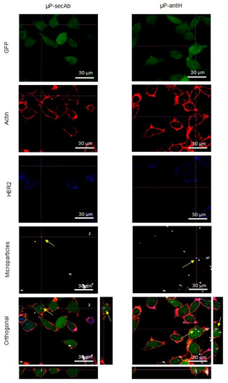
Immunofluorescence analysis by confocal laser scanning microscope (CLSM) of cells cultured in static conditions. Confocal images of D492 and D492HER2 cells cocultured in static conditions and incubated with microparticles biofunctionalized with a non-specific secondary antibody (µP-secAb) or a specific anti-HER2 antibody (µP-antiH). Cells, constitutively expressing green fluorescent protein (GFP, green), were incubated with Alexa Fluor® 546 Phalloidin (red) to label actin microfilaments and Alexa Fluor® 405 conjugate secondary antibody (blue) to label HER2 in the plasma membrane. The arrows point to some examples of µPs located inside the cells. From ‘Cell Internalization in Fluidic Culture Conditions Is Improved When Microparticles Are Specifically Targeted to the Human Epidermal Growth Factor Receptor 2 (HER2).’
“During this process, it was shown that a key parameter, i.e., the spreading angle of atomization could provide a linkage among the working process, the property of generated nanoparticles and their functional performance,” stated the researchers. “They confirmed that the nanoparticle diameter (prepared based on a modified technique) has a profound influence on the drug release performance.”
Shah et al. [5] contributed a study as they moved forward to refine the effectiveness of moxifloxacin, an antibiotic used to treat a variety of bacterial infections. With a new system for optimizing nanoemulsion, they were able to ‘enhance the therapeutic effects of moxifloxacin,’ resulting in safe delivery.
Another development, contributed by Wan et al. [6], allowed for sustained release of loxoprofen sodium (LXP), with the addition of a unique coating:
“Their results identified both the citric acid (CA) and ADEC as the dissolution and diffusion-rate controlling materials significantly decreasing the drug release rate,” stated the researchers. “The optimal formulation for a pH-independent drug release in media has been suggested as at a pH above 4.5 and at slightly slow release in acid medium.”
“The pharmacokinetic studies have revealed that a more stable and prolonged plasma drug concentration profile of the optimal pellets has been achieved, with a relative bioavailability of 87.16% compared with the conventional tablets.”
Studying magnetic nanoparticles, Iglesias et al. [7] stated that BMNPs (biometic) were superior to MNPs (inorganic) for drug loading of molecules ‘positively charged at neutral pH.’ MNPs, however, have the potential for suitable transport abilities while under a magnetic field. Savin et al [8] explored gel formulations for skin melanoma treatment, along with fabricating breast cancer cells in 3D models—following one of the major trends in medicine today using models for treatment, training, and surgical planning too.
“The in vitro results for the tested CD-NHF-loaded gel formulations have revealed that the new composites can affect the number, size, and cellular organization of spheroids and impact individual tumor cell ability to proliferate and aggregate in spheroids,” stated the researchers.
Guadarrama-Acevedo et al. [9] created a new wound dressing made of alginate membrane and polycaprolactone nanoparticles, loaded with curcumin for healing. Integrating nanocarriers allows for drug permeation into multiple layers of skin, thus eliminating solubility issues with curcumin. Rancan et al. [10] performed research showing that after six hours, nanofiber mats offer the best drug concentration upon delivery.
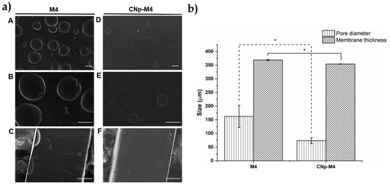
Morphology and porosity of alginate membranes. (a) Micrographs by scanning electronic microscopy of the alginate membrane surface (A, B, D, and E) and membrane thickness (C and F). Magnification of 100× for A, D; 220× in B, C, E and F; the scale bar is 100 μm; (b) pore diameter and membrane thickness of M4 and CNp‒M4 membranes, mean ± SE, n = 3. * indicates that p < 0.05 is statistically significant. From ‘Development and Evaluation of Alginate Membranes with Curcumin-Loaded Nanoparticles for Potential Wound-Healing Applications.’
Further, Lian et al. [11] developed a system for using red blood cell membrane-camouflaged ATO-loaded sodium alginate nanoparticles (RBCM-SA-ATO-NPs, RSANs) to eliminate toxicity of ATO. Their work also showed that RSANS have lower levels of cytotoxicity, upon comparison with normal cells; RSANS also displayed antitumor effects on NB4 cells and 7721 cells.
Adeleke et al. [12] developed an isoniazid suspension as an antitubercular agent against TB, offering extended release:
RDS has been dispersible and stable in the dried and reconstituted states over 4 months and 11 days respectively, under common storage conditions.

Representative graphs displaying: (a) particle size distribution, (b) zeta potential distribution, as well as TEM micrographs showing different surface topographies and characteristics of the reconstitutable dry suspension (RDS) particles at different scales: (c) 1μm and (d) 5 μm, respectively. ‘Development and Evaluation of a Reconstitutable Dry Suspension Containing Isoniazid for Flexible Pediatric Dosing.’
“The published papers are also being compiled as an edited e-book, to be published by MDPI,” stated the researchers.
What do you think of this news? Let us know your thoughts! Join the discussion of this and other 3D printing topics at 3DPrintBoard.com.
[Source / Images: ‘Pharmaceutical Particulates and Membranes for the Delivery of Drugs and Bioactive Moleclules’]
The post 3D Printing Review in Drug Delivery Systems: Pharmaceutical Particulates and Membranes appeared first on 3DPrint.com | The Voice of 3D Printing / Additive Manufacturing.
Recycling Plastics in Fabrication & Design: 3D Printing Raw Materials from Plastic Waste
Alaeddine Oussai, Zoltán Bártfai, László Kátai, and István Szalkai explore ways to improve recycling in 3D printing, releasing the details of their recent study in ‘Development of 3D Printing Raw Materials from Plastic Waste.’
Using plastic and dealing with the trash it produces has become normal around the world—leaving many to look back on the years when we enjoyed products in reusable glass containers—like milk, for example. The authors point out that while plastic waste, regarding type and quality, varies around the world, many types are suitable for re-use. For 3D printing, however, recycling processes are still being heavily explored:
“The current applications for using recycled plastics in fabrication and design are fairly limited, on a small scale, plastics (such as ABS, HDPE, or PET) are shredded and formed into pellets, and then either extruded into lament to be used in existing 3D printers, or injection molded into small parts and pieces of larger components.”
“At a large scale, recycled HDPE is melted into sheets and either used directly as sheets in construction, or then heat formed from a sheet into components for construction. These methods of fabrication using recycled plastics are the norm because of their affordability and straightforward processes, yet each method leaves some complexity to be desired.”
For this study, the researchers focused on both polyethylene terephthalate (PET), often referred to as polyester, and polylactic acid (PLA). While it is true that recycling can be both complicated and confusing for consumers everywhere, there are four basic exercises associated with the process:
- Mechanical reprocessing into equivalent products
- Secondary processing into products with ‘lower properties’
- Chemical constituent recovery
- Quaternary energy recovery
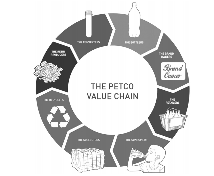
PET recycling circle
Known as a #1 recycling plastic, PET is made of monomer ethylene terephthalate. This is a material often used to make plastic bottles, as well as thin films and solar cells, offering high mechanical strength and temperature resistance.
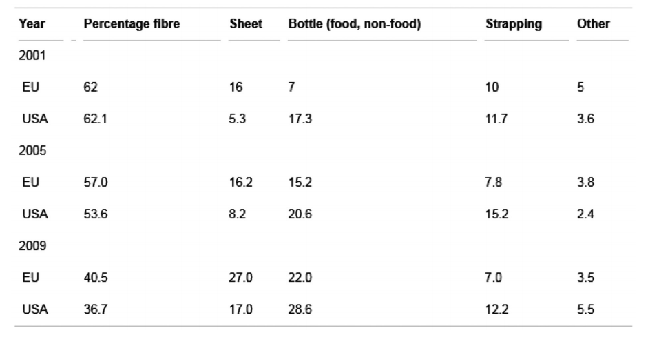
End markets for PET recirculates in Europe and in the USA (Petcore, 2011; Napcor, 2011).
PLA is one of the most common types of 3D printing filament, known for its plant-based nature and resulting biodegradability. And while its true nature in terms of degrading are still being explored, this material is used widely by users and researchers too, featured in studies exploring the potential for a more circular economy, development of biodegradable blends, direct waste printing with pellets, and much more.
“Although PLA is a biodegradable material, which would significantly reduce environmental pollution associated with its waste, the knowledge behind this material recycling and changes in the properties of PLA upon its multiple processing is a very important subject of discussion,” state the authors.
Other materials like polyvinyl chloride (PVC) can break down in only several days. PVC is also affordable and offers the potential for making high-performance parts. Polyethylene’s PE’s are also commonly used, offering density, toughness, and flexibility.
Today, overall recycling efforts, and within 3D printing also, are geared toward less use of energy, less sorting during the process, and the eventual inclusion of what are now non-recyclables. Currently only poly (ethylene terephthalate) (PET) and polyethylene (as 9 and 37% of the annual plastic produced) are being mechanically processed.
The researchers mention a previous research solution as they developed a ‘crusher,’ for re-using plastic. The device consists of a crusher and extruder, along with 12 blades.

overview of the complete design of the crusher

Overview of the complete design of the extruder
Other recycling methods include chemical technology; however, the researchers point out that right it is considered to be too expensive due to the amount of energy it consumes. They point out that incineration is an option too.
“In the current scenario there is growing demand and interest towards chemical recycling methods with low energy demand along with compatibility of mixed plastic waste to overcome the need for sorting and expanding the recycling technologies to traditional non-recyclable polymers,” concluded the researchers.
What do you think of this news? Let us know your thoughts; join the discussion of this and other 3D printing topics at 3DPrintBoard.com.
[Source / Images: ‘Development of 3D Printing Raw Materials from Plastic Waste’]
The post Recycling Plastics in Fabrication & Design: 3D Printing Raw Materials from Plastic Waste appeared first on 3DPrint.com | The Voice of 3D Printing / Additive Manufacturing.
How to Accurately Price for Stereolithography (SLA) 3D Printing Projects
An in-depth pricing guide for 3D printing business and fabrication shops. By Mike Moceri – Founder & CEO MakerOS SLA 3D printing, and all of its variations, offers incredible accuracy for highly detailed parts like electronics enclosures and small miniatures. But so much accuracy comes with an equal amount of complexity and cost. I’ve discovered […]
3D Printing and COVID-19, May 25, 2020 Update: DSM, Amazon, Fortify
Companies, organizations and individuals continue to attempt to lend support to the COVID-19 pandemic supply effort. We will be providing regular updates about these initiatives where necessary in an attempt to ensure that the 3D printing community is aware of what is being done, what can be done and what shouldn’t be done to provide coronavirus aid.
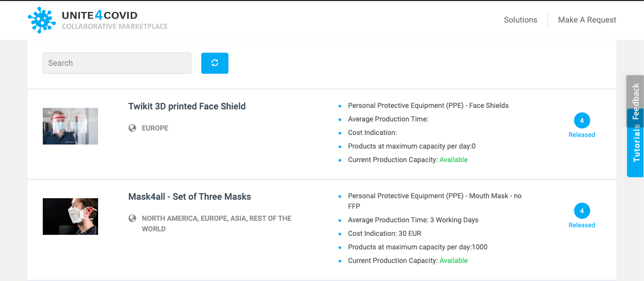
Dutch chemical company DSM has launched a platform for connecting healthcare providers and businesses for the supply of personal protection equipment (PPE) and other items. UNITE4COVID acts as a hub in which manufacturers and certifications labs can be linked to medical professionals in order to provide PPE and safety equipment.
Fortify is 3D printing tooling for injection molding meant to produce an adapter that converts a snorkel into a face mask. By making the tooling with its own 3D printing technology, the company believes that it could reduce the lead time for mold production by 75 percent, cutting a 14-day lead time to three days and potentially reducing costs from $2,000 to $300.
Origin has begun shipping its nasopharyngeal swabs, which are now FDA-registered, Class I, 510K medical products. Bulk orders of the swabs can be ordered on the company’s website.
A thirteen-year-old Tennessee student is using his own desktop 3D printer to produce ear savers, meant to reduce chafing caused by facial masks. Printing at a rate of eight ear savers in three-and-a-half hours, Sam Walker is running his printer 17 hours a day, giving them to local doctors, nurses, pharmacists and retirement home employees.
Also making ear savers is a collective of companies including Ultimaker, HP and other businesses, such as ImageNet Consulting, which has made over 10,000 such devices. While we have covered many of HP’s initiatives so far, Ultimaker’s involvement is made up of a two-part endeavor in which hospitals with pre-approved designs and material specifications are connected to 3D printing companies like 3D Hubs and ImageNet, a substantial U.S. reseller of IT technology and 3D printers, to produce the equipment.
Members of Amazon’s Prime Air mechanical design and hardware teams have joined a Washington State initiative that is producing face shields for healthcare workers. Based on feedback from medical professionals, the Amazon team claims to have improved the initiative’s existing face shield design, including material quality, so that they can be reusable and keep the shield in place more effectively. The new design also reduced sharp edges and pressure to the forehead, as well as improved print time. The Prime Air team subsequently made the U.S National Institutes of Health-approved design available for 3D printing and injection molding.
So far, Amazon has donated about 10,000 face shields and aims to deliver 20,000 more, using systems typically used to cut fiber materials for drone manufacturing to cut the face shield screens. Soon, the retail behemoth aims to mass produce face shields, suggesting it will be able to make hundreds of thousands over the next few weeks and have them available through the Amazon website. A recent blog post on the topic states:
“Because of the design innovations and the capabilities of our supply chain, we are confident we will be able to list them at a significantly lower price—almost a third of the cost—than all other reusable face shields currently available to frontline workers. We are looking to prioritize frontline workers and then eventually open up to all Amazon customers.”
During the pandemic, Amazon has faced harsh criticism from employees, including at Amazon warehouses and Whole Foods, who striking for better conditions as they continue to work amid the health crisis. While warehouse workers in multiple cities are demanding that Amazon shut down facilities where employees have been tested positive for COVID-19, CEO Jeff Bezos has grown his wealth by $25 billion since January 1, which the Institute for Policy Studies has described as “unprecedented in modern financial history.”
A leaked memo indicated that, after firing a worker who played a role in organizing a strike at a Staten Island distribution warehouse, company executives would discredit the worker and the larger movement to unionize Amazon workers. The employee was described as “not smart or articulate.”
According to VICE, who received the leaked documents, the company has attempted to cover its labor record in the midst of the pandemic with public relations efforts. Included in those efforts is the possibility of making free masks, with Amazon General Counsel David Zapolsky saying that the corporation should come up with “different and bold” ways for giving away surplus masks.
3D-printed medical supplies have obviously generated a lot of positive PR for companies involved in producing them. What this recent news could indicate is that the move by Amazon to participate in making 3D-printed face shields is part of a larger campaign to generate good publicity in the face of its labor disputes. Because we have seen multiple other companies laying off workers also participate in the production and delivery of medical supplies, such as GE and Boeing, it might not be unreasonable to think that similar campaigns are under way by other entities.
As the pandemic continues to grip the world, we will continue to provide regular updates about what the 3D printing community is doing in response. As always, it is important to keep safety in mind, remain critical about the potential marketing and financial interests behind seemingly good humanitarian efforts from businesses, and to do no harm.
The post 3D Printing and COVID-19, May 25, 2020 Update: DSM, Amazon, Fortify appeared first on 3DPrint.com | The Voice of 3D Printing / Additive Manufacturing.
3DHEALS2020: A Not So Lonely Planet
Only a few weeks away from 3DHEALS2020, and I just got off the phone with one of our speakers, Dr. Ho, from NAMIC Singapore. Our brief interview reminded me just how much I enjoyed Singapore—its start-up like government, incredible universities, and its beautiful modern architecture, chili crabs, and unpredictable rainstorms. Now, I’m on my way to some of the best meals in my life with another 3DHEALS community event in a foreign city. Looking back, there are many stories like that: in Detroit, Vigo, Paris, Shanghai, or Boston, my work with 3DHEALS communities has been a journey of adventures and friendships. 3DHEALS2020 is really a way to summarize my travels from the last two years. It is my version of Lonely Planet—the healthcare 3D printing version.
I really felt more alive when people have welcomed me into their cities; when they have showed off their latest innovations; when they have bantered enthusiastically with one another in a local pub till midnight after 3DHEALS events. And they felt the same way.
Sadly, however, this pandemic is putting old methods of human connection into question. Perhaps, a virtual summit is a stopgap solution for conferences, but, more likely, it is time for us to explore alternative and better ways to stay connected and informed.
The virtual 3DHEALS2020 summit will be a good start.
While we can’t serve you delicious San Francisco Blue Bottle coffee, there are three things we aim to do right with this conference:
1. Awesome live content
One upside about the virtual summit is that people who could not be available due to logistic barriers are now more available. We added 20+ speakers since the pandemic began and are still adding more parallel workshops to the existing program. Some of highlights include:
A. Biofabrication/Bioprinting Panels and Workshops:
Welcome to the holy grail of healthcare 3D printing applications!
These panels and workshops collect some of the brightest minds in the world of tissue engineering, biofabrication, and bioprinting. It includes the newest generation of startup founders. Names such as Stephanie Willerth, Adam Feinberg, Jordan Miller are already well-established and loved in the scientific communities and just founded their own startups within the last 12 months. More established startup founders whose companies are also critical to the eventual success of biofabrication, tissue engineering, and cell therapy at large will also join us live, including Melanie Mathieu from Prellis Biologics, Jon Rawley from Roosterbio, John O’Reily from Xylyx, Taciana Pereira from Allevi, and Kevin Caldwell from Ossium Health. Qrquidea Garcia (“Orchid”) from JNJ Innovation will also join us on this panel, discussing how an industry leader can work with innovators and startups in this exciting, burgeoning field.

B. Regulatory and Legal Landscape of Healthcare 3D Printing
For those who put their skin in the game, this is probably one of the most must-attend sessions. 3D printing in healthcare is a super new field, and legal experts in this field who have established track records and legitimacy are only a handful. This session will include the most comprehensive list of legal and regulatory concerns specifically for healthcare 3D printing, including intellectual property/patent issues, product liability, FDA pathways, manufacturing standards, and more. Steven Bauer, from FDA CBER, just joined the panel to directly address concerns related to cell therapy from the biofabrication and stem cell communities. The speakers are not just well-versed on how to interpret the law and policies, but also how to interact with scientists, policy makers, organizations, and standards bodies at this early stage of the industry, with practical, real-life examples.

C. Global Perspectives
One lesson from this pandemic is that globalization has consequences. Having a well-rounded worldview of the global healthcare 3D printing ecosystem is a requirement for future success. Our early morning sessions are reserved for international speakers all over the world to meet the audience and share their unique perspectives, needs, and hopes. Both America Makes director John Wilczynski and NAMIC director Dr. Chaw Sing Ho, along with experts from Turkey, India, and Taiwan, will share how healthcare innovations can thrive in both local and global environments. On day two (June 6th), the audience will learn about how different countries are implementing the concept of 3D printing for Point of Care, which cannot be taken out of context of different healthcare systems and cultures. The audience will meet and learn from the leaders at UCSF, Stanford, Germany (Kumovis), India (Anatomiz3D), and developing countries.

2. Pre- and post-event networking opportunities
The attendees will have the opportunity to meet other attendees, speakers, and conference organizers as soon as they sign up the event using a dedicated conference app. They can send direct messages, post threads, share photos, host their own virtual events days before the conference. The app will be available to registered attendees for six months after the conference ends.
3. Entrepreneurship
One of the most exciting aspects of 3DHEALS2020 is its focus on entrepreneurship. Pitch3D has been a quarterly free and online pitch platform to selected early-stage startups in healthcare 3D printing and bioprinting spaces for the last two years, introducing 30+ startups from all over the world to institutional investors. 3DHEALS2020 also gathered some of the most experienced VCs and entrepreneurs in the space to share their stories, perspectives, and directly engage with the startups and the 3DHEALS2020 attendees directly during both pitch sessions and investor panels. There will be ten startups pitching each day at 5-6 PM PST. Interested startups can apply here.
This is the time of uncertainty and change.
Join us at 3DHEALS2020, connect with the world, and take control of your future. This is a Not So Lonely Planet.
The post 3DHEALS2020: A Not So Lonely Planet appeared first on 3DPrint.com | The Voice of 3D Printing / Additive Manufacturing.
Making a Mini Arcade Wireless Charger #NeoPixel #CPX #3DPrinting
YouTube Makers EvanAndKatelyn have fun creating this mini arcade. The charger is taken to the next level with a Circuit Playground and some LEDs.
We made a wireless phone charger that looks like a mini arcade and even has a working button! Click the link to download PUBG Mobile for free: http://www.inflcr.co/SH2Bt. Thanks to PUBG Mobile for sponsoring this video!
A Guide to Bioprinting: Understanding a Booming Industry
The success of bioprinting could become the key enabler that personalized medicine, tissue engineering, and regenerative medicine need to become a part of medical arsenals. Breakthroughs in bioprinting will enable faster and more efficient patient care and recovery. Biofabrication could be used to reshape the foundations of drug development, medicine, cosmetics, organ transplantation, and many other fields. It will transform the way doctors repair damaged ligaments, recreate tissues, and even reproduce the layers of the skin.
We are entering an era of bioprinting revolution. But to understand the role that bioprinting will play in the future, it is important to look back at how early discoveries in the field provided a strong basis to push its capability forward:
- Back in the ’90s, Anthony Atala, pediatric surgeon, urologist, and director of the Wake Forest Institute for Regenerative Medicine (WFIRM) in North Carolina, created by hand bladders, skin, cartilage, urethra, muscle, and vaginal organs. By the end of that decade, the Institute used a 3D printer to build a synthetic scaffold of a human bladder, which they then coated with cells taken from their patients and implanted it, preparing the stage for bioprinting.
- Then in 2002, scientists from Harvard University printed two-inch-long mini-kidneys capable of filtering blood that was then transplanted back into genetically identical cows, where they started making urine. The novel research raised the prospect of using stem cells taken from human patients with kidney failure to create new organs for transplant.
- However, it wasn’t until 2003, when bioengineering professor Thomas Boland adapted a Hewlett-Packard inkjet printer in his lab at Clemson University to begin printing a bioink made of living bovine cells suspended in the cell-culture medium, that bioprinting began to materialize. This led to the creation of the world’s first 3D bioprinter, capable of creating living tissue from a solution of cells, nutrients, and other bio-compatible substances.
A member of WFIRM team operates with the bioprinter (Image: WFIRM)
Twenty years later, researchers still face challenges as they continue working with bioprinters and bioinks. Even though there has been an increasing adoption of the technology, the extent of its potential has not been fully exploited. From choosing the bioinks to actually bioprinting human tissues and organs, this new field is quickly becoming the go-to technology that bioengineers, researchers, and hospitals need to evolve from lengthy and cumbersome manual work to scalable and replicable results.
How Well Do you Know Bioprinters? (or How Bioprinters Work and Who Makes Them)
Bioprinters work by extruding cells and other biomaterials contained in bioinks, from syringes that deposit the material layer by layer to create different types of tissues or organ-like constructs. The technology behind the bioprinters vary. Nonetheless, to date, the three main and most popular bioprinting technologies are extrusion, inkjet, and laser-based bioprinting. Some mainstream examples are:
- Some manufacturers, like Cellink or Allevi, use pneumatic-driven extrusion systems that pump high-pressure air in a cartridge to force bioinks to flow through a nozzle.
- Other fabrication systems, such as the one designed by Poietis has laser-assisted bioprinting that allows cells to be positioned in three dimensions with micrometric resolution and precision to design living tissue.
- Another type of bioprinting technology uses a stereolithography-based bioprinting platform. Vendors using this process include Volumetric and Cellink’s jointly produced Lumen X projection stereolithography based bioprinter.
- Another project that could revolutionize the way surgical procedures are performed is handheld bioprinters; these systems enable surgeons to deploy cells — or material to aid in cellular growth — directly into a defect site in the body, such as severely burnt skin, corneal ulcerations or bone. One of the most talked-about handheld bioprinters has been Australia’s University of Wollongong BioPen, allow surgeons to repair damaged bone and cartilage by “drawing” new cells directly onto bone in the middle of a surgical procedure. Although still in pre-clinical trials, these devices have attracted the attention of healthcare practitioners due to its versatility.
A few of the main manufacturers supplying the market include 3D Bioprinting Solutions, Allevi, Aspect Biosystems, Cellink, nScrypt, regenHu, Inventia, Regemat3D, Poietis, and more. Last year, 3DPrint.com counted 111 established bioprinting firms around the world. Mapping the companies that make up this industry is a good starting point to understand the bioprinting ecosystem, determine where most companies have established their headquarters and learn more about potential hubs, like the one in San Francisco.
Types of Bioinks
3D bioprinters use bioinks. Bioinks are substances made of living cells that can be used for 3D printing complex tissue models — they mimic an extracellular matrix environment to support the adhesion, proliferation, and differentiation of living cells. Choosing which bioink to use can be challenging. To date, we have witnessed researchers using bioinks based on several biomaterials, such as alginate, gelatin, collagen, silk, hyaluronic acid, even some synthetic-biomaterials-based-bioinks.
The promise of hydrogels. A macromolecular polymer gel constructed of a network of cross-linked polymer chains, hydrogels are able to meet the stringent requirements of cells and are the basis of almost all bioink formulations. As stated in “Engineering Hydrogels for Biofabrication”, published in Advanced Materials, “hydrogels are particularly attractive for biofabrication as they recapitulate several features of the natural extracellular matrix and allow cell encapsulation in a highly hydrated mechanically supportive three‐dimensional environment.” This makes hydrogel-based bioinks a very promising choice for many researchers and bioengineers.
Bioinks from patients’ cells. The biomaterials can also use a patient’s own cells, adult stem cells, manipulating them to recreate the required tissue. The source of the cells varies depending on what researchers are bioprinting. For example, in the case of Alzheimer’s disease, experts at the University of Victoria in Canada, have bioprinted neural tissues using stem cells as a tool for screening drug targets for the disease. The ability to program patient-specific cells is the beginning of customized bioprinting since the unlimited potential of these cells can be used to regenerate or repair damaged tissue.
Polbionica’s bioinks (Image: Polbionica)
What is Bioprinting Good For?
What is most exciting about bioprinting, are the many ways that doctors and researchers are using currently available devices in the market or are creating their own systems to facilitate new processes and applications. The orchestrated interaction between machine and user has led to innovation that could reinvent the world of tissue engineering.
Moving oncology forward: Bioprinting is being employed in the battle against cancer, whereby scientists create tumor models for research. Modeling cancer using 2D cell cultures fails to accurately replicate the microenvironment of tumors. This is why scientists have turned to biofabrication tools to make three-dimensional models that mimic the intricate in vivo tumors. These models help test anticancer drugs; aid scientists in understanding the underlying causes of metastasis, and can even personalize treatment for individual cancer patients. There have been plenty of initiatives that apply bioprinting to oncology. These range from immersion bioprinting of human organoids to printing cancer tissues in 3D.
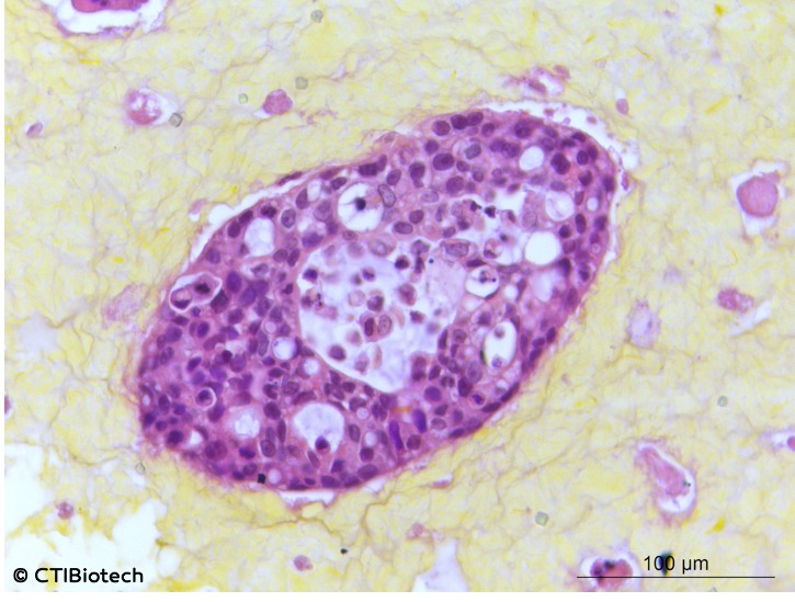
Microtumors (Image: CTI Biotech)
The market for oncology-oriented bioprinting seems sure to grow. The number of patients suffering from the disease continues to go up. In 2018 alone there were 17 million new cases of cancer worldwide, and projections suggest that there will be 27.5 million new cases of cancer each year by 2040. What that effectively means is that we are witnessing an increase in oncology bioengineering research and whether it is for glioblastoma, bone cancer tumors, or lung models with tumors, the implications can be profound since the ability to use bioinks and bioprinters to create tumors frees researchers of the many ethical concerns associated with testing as well as reduces the costs associated with such research activities.
Building scaffolds: Probably the most important practical use for bioprinting at the present time is in regenerative medicine. For instance, in 2019, researchers from North Carolina State University (NCSU) and the University of North Carolina at Chapel Hill created a 3D biomedical fiber printer used to create biocompatible scaffolds. Also, Harvard researchers working in Jennifer Lewis’ Lab at Harvard´s Wyss Institute for Biologically Inspired Engineering and John A. Paulson School of Engineering and Applied Sciences (SEAS), came up with a much talked about breakthrough new technique, the SWIFT method, that allows 3D printing to focus on creating the vessels necessary to support a living tissue construct. A team of researchers at Texas A&M University have even developed a 3D printable hydrogel bioink containing mineral nanoparticles that can deliver protein therapeutics to control cell behavior.
Limitations of Bioprinting
Though we heard it many times before, knowing that someday 3D printed artificial organs could eliminate the need for an organ donor waiting list is comforting. Creating personalized replacement organs sounds like the solve-all solution to the organ shortage crisis, yet, a functional organ compatible for human implantation may be decades away. Today, 3D printed organs are still raw to be used for transplantation and lack the vasculature required to function within the human body.
Creation. Last year, mainstream news outlets headlined a story about researchers who had 3D printed a heart. However, the published scientific paper behind that story described how a group of scientists from Tel Aviv, Israel, created bioink out of heart cells and other materials from a patient, and were able to develop cardiac patches and ultimately, 3D print comprehensive tissue structures that include whole hearts. The tissue was shaped in the form of a tiny heart that was kept alive in a nutrient solution. The paper expresses how this development could not function like a real heart since the cells in the construct can contract, but don’t yet have the ability to pump.
This was certainly not the first heart to be 3D printed, yet Tal Dvir, who led the project at Tel Aviv University’s School of Molecular Cell Biology and Biotechnology, indicated that never before had it resulted in an organ “with cells or with blood vessels.” It was an amazing breakthrough for the field, and it proves that biotechnology has made significant advances, but it is still a long way from creating organs that can be transplanted to people, considering that the vasculature — the network of blood vessels that feeds the organ — remains a challenge, but scientists are determined to troubleshoot these issues.
So, no matter how enticing the idea of successfully bioprinted organs sound, stories like this remind us to keep the hype in check, making the work of news outlets fundamental for reporting advancements in research and medical breakthroughs (which usually take much more time).
Where to Next?
Organ bioprinting. The application of 3D bioprinting will be a game-changer in medicine, as the machines successfully replicate tissues and organs, build muscles and cartilage, and enable the adoption of customized medicine. The long-term dream for bioprinting has always been the routine printing of body organs. Current ongoing projects include Michal Wszola’s 3D bioprinted bionic pancreas or Organovo’s 3D bioprinted liver.
The space frontier. Bioprinting in space could hold the key to developing fully functional organs. This is because bioprinting without gravity allows organs to grow without the need for scaffolds. The National Aeronautics and Space Administration (NASA) considers that terrestrial gravitation represents a significant limitation, while a gravity-free environment, magnetic and diamagnetic levitation will allow for biofabrication of 3D tissue constructs with a scaffold-free and even nozzle-free approach. Some bioprinters have already been launched and used in space. This includes nScrypt and Techshot’s BioFabrication Facility (BFF), or the Organaut 3D bioprinter created by Russian biotech firm 3D Bioprinting Solutions and Roscosmos, the Russian state corporation responsible for space flights.
Healthcare at its best. More prosaically, biomaterials specialist and a professor of biofabrication at Queensland University of Technology, Australia, Mia Woodruff, has been advocating the hospitals of the future for years. She has an exciting vision of a future where the fabrication of patient-specific replacement tissue and organs is safe, cost-effective, and routine. Though perhaps years from happening, her vision is in tune with what many think bioprinting could become, that is, with enough researchers, companies, and funding.
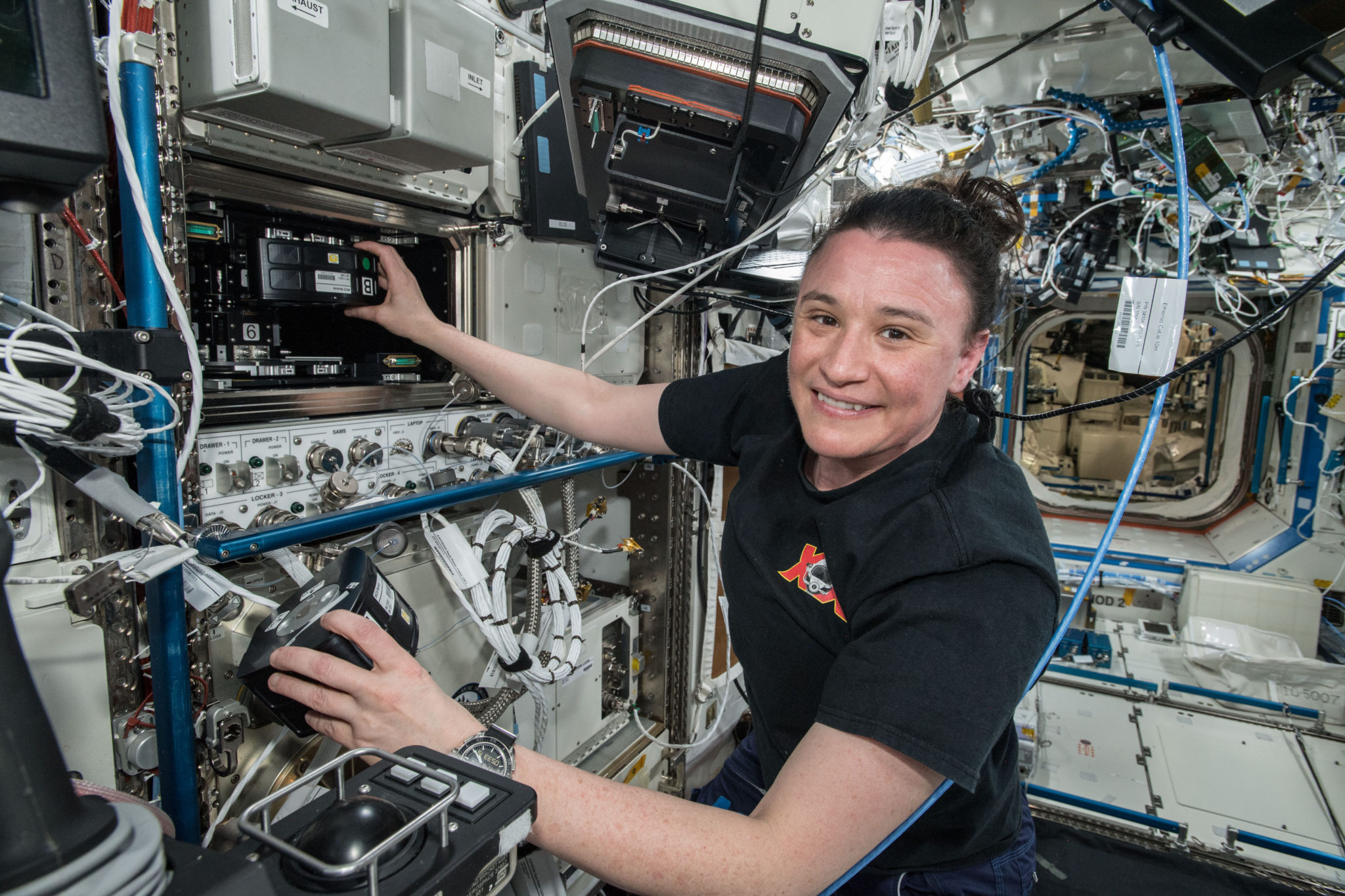
An astronaut aboard the ISS using Techshot’s BioFabrication Facility or BFF (Image: Techshot/NASA)
Coming regulation. Back here on Earth, there will be a growing need for common guidelines for bioprinting to make the process more standardized. In the EU, for example, there currently no particular regulatory regime governing the whole bioprinting process, but piecemeal legislation is relevant in relation to tissue engineering and regenerative medicine. While the Food and Drug Administration (FDA) of the United States plans to review the regulatory issues related to the bioprinting of biological, cellular and tissue-based products in order to determine whether additional guidance is needed beyond the recently released regulatory framework on regenerative medicine medical products.
As the development of the technology strongly advances and proves successful for researchers, we will surely continue to observe brilliant minds perfecting devices and biomaterials, envisioning new systems for future needs, especially as startups emerge out of universities and research institutes, and established companies upgrade their machines to face the limitations we previously addressed in this article.
Still, there is a long way to go, what was largely built so far is a very promising technology. For instance, fully functional organ fabrication for transplantation might take decades. Nonetheless, the unquestionable contribution of bioprinting to so many fields remains an incentive to invest in this area to overcome medical challenges and to move the healthcare industry in a different path, where technology will not only aid in curing diseases but also guiding people by helping them stay healthy, recognizing symptoms early and personalizing solutions in real-time.
The post A Guide to Bioprinting: Understanding a Booming Industry appeared first on 3DPrint.com | The Voice of 3D Printing / Additive Manufacturing.
Cell Culture Bioreactor for Tissue Engineering
Researchers from the US and Portugal are refining tissue engineering applications further, releasing the findings of their study in the recently published ‘A Multimodal Stimulation Cell Culture Bioreactor for Tissue Engineering: A Numerical Modelling Approach.’ In creating a new bioreactor for 3D printing, the authors worked to promote reproducibility and optimization, fabricating a design not possible using conventional techniques.
In most bioprinting, cells are seeded onto scaffolds—also the source of much study, whether in regard to new techniques, enhancements, or interface engineering—and then researchers must hope for viability. Promoting cell growth and sustainability is one of the greatest challenges in any type of bioprinting, and often devices like perfusion flow bioreactors are used in replacing culture mediums to remove toxins. In some cases, they are also used for mechanical stimulation through fluid flow shear stress (FFSS). Electric field stimulation devices can also be easily used to encourage cells to grow, mature, and differentiate.
The bioreactor used here was part of a previous study conducted by the authors, but now updated (using SOLIDWORKS 2018 Student Edition ) to include capabilities in E-Field stimulation and fluid flow mechanical stimulation. 3D design was performed in SOLIDWORKS 2018 Student Edition. Assessment of the new design was part of this study, leaving the team to create a numerical finite element analysis (FEA) of the model.
With FEA, the researchers could project input conditions for the bioreactor, further enhancing effectiveness of cell stimulation, determined from in vitro data. Overall, in vitro tests can offer ‘essential’ information for confirming ranges of multimodal stimulation—projected via numerical studies.
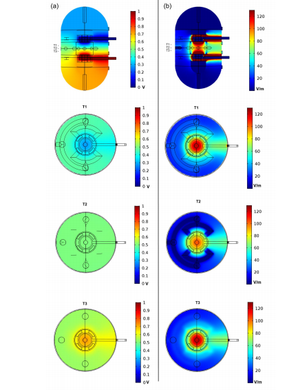
Numerical finite element analysis (FEA) analysis of the proposed bioreactor design with a DC electric stimulation parallel plate capacitor set-up with lateral and top slice views. The three top views represent the ROI upper slice (T1), the ROI middle plane slice (T2) and the ROI bottom slice (T3). (a) Electric potential distribution predicted in the bioreactor due to DC stimulation. (b) E-Field magnitude distribution predicted for the same electric DC stimulation conditions.
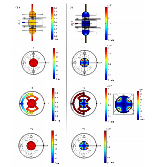
Numerical FEA analysis of the proposed bioreactor design for a laminar perfusion flow with lateral and top slice views. The three top views represent the ROI upper slice (T1), the ROI middle plane slice (T2) and the ROI bottom slice (T3). (a) Pressure distribution predicted considering applied inlets velocity of 0.003 m/s and a outlet pressure of 0 Pa. (b) Fluid velocity distribution predicted for the same inlet/outlet conditions. The velocity distribution at the ROI middle plane slice is presented in more detail in a top view inset at the right of the slice plane.
“Electrical and mechanical stimulation conditions in the region-of-interest (ROI) were considered for bone cell stimulation optimization, according with reference values obtained from two previous in vitro studies on bone cell stimulation, one applying mechanical stimulation, and the other using E-Field stimulation,” explained the authors.
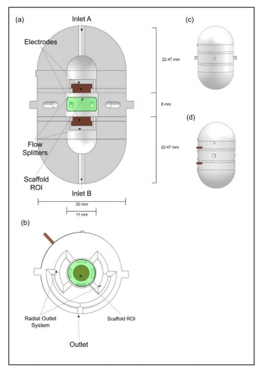
Novel bioreactor design: (a) Vertical cut view of the bioreactor design, where the parallel electrodes set up, the upper and bottom inlets and the inlet flow splitters can be observed. (b) Horizontal cut view of the bioreactor design, where the radial outlet system can be observed. The green regions represent the region-of-interest (ROI) where the scaffold will be placed, represented by a cylinder with 4 mm of height and a diameter of 10 mm. (c) CAD bioreactor design assembled in frontal view, the main outlet hole is visible in the middle. (d) CAD bioreactor design assembled in lateral view, showing both electrode connector wires (in brown).
Flow splitters were added between the inlet and the scaffold, resulting in ‘indirect flow prevalence.’ Inlets and outlets were fitted with hose joiners, connecting them to the perfusion pump. The cell culture chamber was separated into two different areas for cell cultures and cell seeding exercises. Materials for use in the platform were required to be non-toxic and suitable according to ISO 10993-5 standards.

Bioreactor geometry volume mesh created using COMSOL Multiphysics, with 1.9 × 10⁶ elements, and an average element quality of 0.65.
“Accordingly, in the direct contact test, cells cultured in contact with all the materials presented normal fibroblast morphology with no evidence of any inhibition halo effect or cell death. According to the cytotoxicity tests results, all candidate materials are suitable for our bioreactor AM fabrication,” concluded the researchers. “We will consider C8 and PETG as materials of interest for future design fabrication. C8 is a new material with good layer adhesion and surface quality, which are key features for the perfusion flow. The C8 supplier datasheet reveals that this material has a higher tensile strength than ABS, resulting in improved mechanical characteristics, which are important for the overall robustness of the bioreactor to withstand the tightness of pressure chambers.”
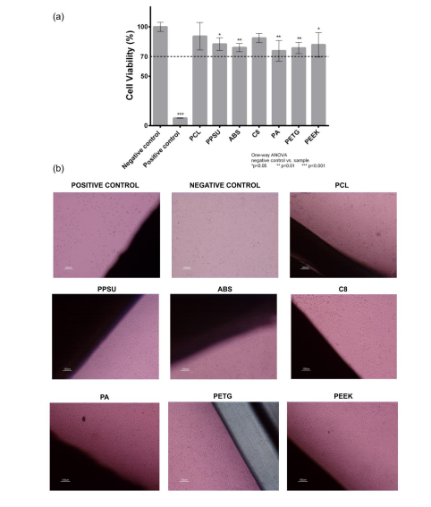
Cytotoxicity assay with L929 mouse fibroblast according to ISO 10993-5 standards: (a) indirect contact (MTT protocol); (b) direct contact (digital images of the material samples and the negative and positive controls, fresh culture medium and Latex, respectively). A one-way ANOVA with no corrections for multiple comparisons (Fisher’s test) statistical analysis was performed using GraphPad Prism6.
“A design–numerical modelling approach will be essential to understand the underlying biophysical effects of electric and mechanical stimuli in cell cultures and can be a powerful tool for standardization of stimulation protocols considering different bioreactor designs and specific TE outcomes.”
What do you think of this news? Let us know your thoughts! Join the discussion of this and other 3D printing topics at 3DPrintBoard.com.
[Source / Images: ‘A Multimodal Stimulation Cell Culture Bioreactor for Tissue Engineering: A Numerical Modelling Approach’]
The post Cell Culture Bioreactor for Tissue Engineering appeared first on 3DPrint.com | The Voice of 3D Printing / Additive Manufacturing.
Korean researchers investigate the effect of temperature on particle emission rates during FFF
A recent study by researchers from Seoul National University investigates the effect of nozzle temperature on the emission rates of hazardous particles during FFF 3D printing. The scientists tested four different common polymer filaments and measured the concentration of particles with direct-reading instruments at various temperature intervals. The work aims to demonstrate that nozzle temperature […]