3D Printed Flow Straighteners for Better Management of Aerosol Particles

In the recently published ‘Improved performance of Differential Mobility Analyzers with 3D-printed flow straighteners,’ researchers from Cyprus and the Netherlands evaluated the development of a customized DMA meant to further streamline building enhanced flow straightening systems for aerosol management.

Combining both flow and an electrostatic field for classification, the DMA is a critical measurement tool for aerosol particles, whether at the nanometer or micron. Flow fields can be difficult to create due to the merging of two flows, often causing issues such as local eddies that may result in diminished performance. In attempting to overcome challenges, aerosol flow may be maintained ‘lower than ca. 10% of the sheath flow.’ Flow straighteners can also be made from a fabric mesh insulator that may cause disturbances but also prevent development of a comprehensive velocity profile.

In this study, the researchers fabricated nine different FSs samples, evaluating performance with a new 3D printed cylindrical DMA. Four were printed with ABS, and the others were made of small nylon meshes, to include Dacron (used as a reference).

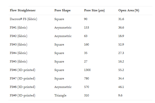
Characteristic details of all the tested FSs.

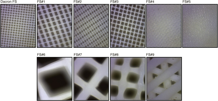
Images of the tested flow straighteners determined by optical microscopy.

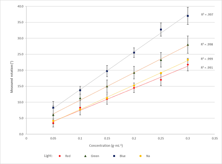
“The performance of each FS was assessed by experimentally determining the Transfer Function (TF) of the DMA (i.e., the function giving the probability of a particle of a given electrical mobility, or size, that enters the instrument, to exit through its monodisperse particle outlet) when that was operated under different operating conditions,” stated the authors.

TF height corresponded to the greatest probability of particles entering the DMA, then exiting via the mono-disperse particle outlet. Deviations were measured in terms of height, with FWHM calculated for each FS and each sheath flow rate.

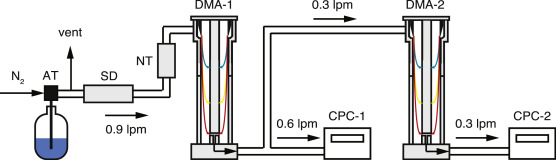
Schematic layout of the experimental set-up used to determine the performance of the test DMA (i.e., DMA-2) when different FS were employed. Key: AT: Atomizer; SD: Silica-gel diffusion drier; NT: Aerosol charge neutralizer; DMA: Differential Mobility Analyzer; CPC: Condensation Particle Counter.

Tests were performed with the following details:

  • Use of nearly monodisperse particles, with diameter of ca. 50 nm
  • Parametric version of the TF captured deviations between both measurements and predictions
  • Fitting parameters affected TF regarding height and width

“In each TDMA experiment we obtain a set of N1 and N2 values, corresponding to the particle number concentrations measured downstream of DMA-1 and of DMA-2, respectively,” explained the authors. “Assuming that P0and CPCRatio remain constant throughout each experiment lasting for ca. 20 min, and that the TF of DMA-1 (i.e., Ω1 in Eq. 1) is constant and well-defined, the TF of DMA-2 (i.e., Ω2 in Eq. 1) is obtained by using a custom-made fitting algorithm (cf. supplement).”

“It should be noted here that all FSs tested in this work created a small step (ranging from 0.7 to 1.0 mm in height) between their outer boundary (for the 3D-printed), or rings (for the fabric), and the walls of the inside electrodes of the DMA.”

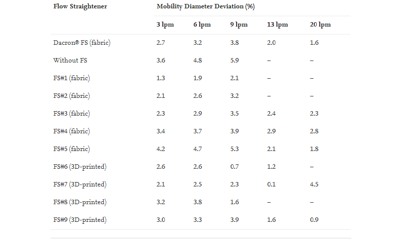
Summary of the results reflecting the sizing accuracy of the test DMA (expressed as % difference between measured and predicted particle size selected by the DMA) when using different FSs.

The researchers followed the Dacron FS used for reference, working to maintain suitable comparability, but noting that because all the samples resulted in steps of similar heights, results were ‘highly intercomparable.’ FS#9 outperformed most of the samples—even the one made from Dacron.

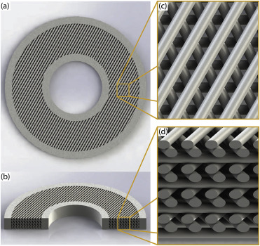
Render illustrations of FS#9, including top view (a), 3D cross-sectional view (b), as well as zoomed in views of the top (c) and of the cross-section (d) of the porous part of the FS.

“The good performance of the DMA when using FS#9, which was better even from the case when the reference Dacron® FS was employed at sheath flow rates up to 20 lpm, warrants for further investigating the use of 3D printed straighteners to run conventional and/or low-cost DMAs (Barmpounis, Maisser, Schmidt-Ott, & Biskos, 2015) at substantially higher sheath flow rates (e.g., up to 20 lpm) or with high-flow DMAs (Fernandez de la Mora & Kozlowski, 2013),” concluded the researchers. “For the latter, which are used for classifying nanoparticles (Wang et al., 2014) and even atomic clusters (Maisser, Barmpounis, Attoui, Biskos, & Schmidt-Ott, 2015) with high resolution, flow laminarization is rather important, typically requiring an additional pre-laminarization stage (Amo-Gonzalez & Perez, 2018).”

“In summary, the measurements reported in this work show that 3D printing can be used to manufacture flow straighteners that exhibit better performance compared to traditional systems made of nylon fabrics. This has been verified by assessing deviations between the measured and the theoretical transfer function of a custom-made DMA employed in the tests. Interestingly, the 3D-printed straightener, having a triangular pore shape combined with a high surface density and larger pores compared to the other FSs investigated here, attributed to the test DMA a performance that compared better with theory. Considering the flexibility and ease of manufacturing of 3D printing, our results show that employing this technology to build flow straightness for DMAs, can improve their performance and thus is a better alternative compared to existing practices.”

What do you think of this news? Let us know your thoughts; join the discussion of this and other 3D printing topics at 3DPrintBoard.com.

[Source / Images: ‘Improved performance of Differential Mobility Analyzers with 3D-printed flow straighteners’]

The post 3D Printed Flow Straighteners for Better Management of Aerosol Particles appeared first on 3DPrint.com | The Voice of 3D Printing / Additive Manufacturing.

Researchers Create Low-Cost 3D Printed Polarimeter for Use in Chemistry Classrooms

The adoption of 3D printing in the classroom has opened up new horizons for creating teaching tools. Science teachers, in particular, can make personalized models of nanostructures, and educational tools like colorimeters. But there haven’t been any 3D printable designs for polarimeters, which measures the angle of rotation of polarized light once it’s passed through an optically active solution or substance. Paweł Bernard from Jagiellonian University and James D. Mendez from Indiana University – Purdue University Columbus published a paper about their creation of a low-cost 3D printed polarimeter.

“3D printing and simple electronics were used to create a polarimeter suitable for a variety of chemistry courses,” they wrote. “This device allows instructors to demonstrate optical activity but is also easy to use and low cost enough to be widely available for student use, as well. The instrument uses an LED light source and detector housed in a 3D-printed base. By rotating the top piece, users can visually detect changes in brightness or measure this directly with a multimeter.”

A polarimeter consists of a sample chamber, monochromatic light, and a polarizing filter before, and a rotatable one behind, the sample. This second filter can be adjusted to the angle of the rotated light, after it’s passed through a sample, in order to “minimize or maximize the transmitted light.”

Basic polarimeter schematic and working theory.

High school and college teachers normally demonstrate the optical activity of substances using overhead projection, as most regular polarimeters are too expensive for use in a school laboratory setting. One researcher created a no-cost polarimeter using sunglasses and a mobile phone, which was good for demonstration purposes, but not for student experiments. Another inexpensive polarimeter was made using a shoebox, but it wasn’t durable enough.

“Therefore, the use of 3D printing technology is a perfect solution,” the researchers stated. “The body of a polarimeter can be printed in a reasonable time; the price of the plastic and electronics is low, and the actual assembly of the elements is relatively simple.”

3D printed polarimeter schematic.

A basic polarimeter can use either a test tube or 3D printed cuvette, and light detection can be merely eyeballed, or precisely measured with a low IR radiation sensitivity photodiode. Both are compatible with low-voltage, inexpensive LEDs; the RBG diode at the bottom can be plugged into a 4.5 or 5 V battery, or a standard 9 V battery can be used with a simple circuit.

9 V power supply circuit schematic.

“In the construction, two layers of polarizing filters (polarizing film) are used. It is a low-cost, commercially available material, used for the construction of 3D glasses among other things,” Bernard and Mendez explain. “Our experience shows that it is easier to identify the lowest (rather than highest) intensity of the light passing through the sample; therefore, we advise arranging two layers of polarizing film rotated by 90°. In such a setup at neutral position (0° angle) without a sample, or with a sample of optically nonactive substance, it is dark, showing the lowest light intensity. 

“The construction of the device using a test tube as a sample container is simpler but also more problematic in use. The bottom of a test tube scatters the light. Usually, the center of the light spot is darker, but there is an unpolarized light ring around it.”

A test tube does not ensure a complete blackout at the minimum light point, so a 3D printed container with a flat bottom is useful. The researchers 3D printed the elements out of ABS and PLA filaments, which were black to ensure stable light readings. PVA supports and a dual extruder printer were used to 3D print the rotary cup and main body.

(a) Operating 3D printed cuvette polarimeter with photodiode detector at zero position (minimum signal); (b) operating 3D printed test tube polarimeter (maximum signal); (c) operating 3D printed test tube polarimeter (min signal); (d) operating the 3D printed cuvette polarimeter (max signal); (e) operating 3D printed cuvette polarimeter (min signal).

The researchers tested 50 high school chemistry students in Poland and 15 organic chemistry university students in the US on taking measurements with the 3D printed polarimeter. Working in groups of 2-3, they ran measurements with pure liquids first, and then aqueous solutions. It’s quick and easy to use – the students can change samples and adjust a cap rotation in less than a minute, though they must be told which way to rotate the tool for different substances as “the device gives the same readings in both directions (90° = −270°).”

“It is also advised to adjust the concentration of the sample solution and path length so that the readings are in the range of the provided rotation scale (from −180° to +180°). Using measured rotation and simple mathematical relations, students can calculate a substance’s specific rotation,” the researchers said.

The students used (R)-limonene, fructose, and sucrose, and ran initial measurements both visually and with the 3D printed polarimeter, which allowed them to take measurements with three colors thanks to its RBG diode. They made 4 to 6 measurements for a sample and after dilation for the aqueous solutions.

“The results were a starting point for a discussion on optical rotatory dispersion phenomena. Calculating the specific rotation of the substances was homework, verified by the teacher during subsequent classes,” the researchers stated.

Measured rotation for aqueous solutions of sucrose in the concentration range of 0.05–0.35 g·mL, a series for red, green, blue light measured with a 3D printed polarimeter, and accompanied by results from commercial polarimeter with a sodium lamp 589 nm.

In another project, instructors prepared kits with all of the materials needed to assemble the polarimeter, including breadboards and the 3D printed body. 16 chemistry majors in Poland and four US undergrad students constructed the device, working in pairs, and none had previous experience using breadboards or building measuring devices. But they followed detailed instructions, with some help from teachers, and succeeded in building operational polarimeters in less than one hour.

Discuss this and other 3D printing topics at 3DPrintBoard.com or share your thoughts below.

The post Researchers Create Low-Cost 3D Printed Polarimeter for Use in Chemistry Classrooms appeared first on 3DPrint.com | The Voice of 3D Printing / Additive Manufacturing.

3D Printing and COVID-19 (Update April 15)

Operation Shields Up OSU covidAs countries around the world grapple with the spread of the coronavirus (COVID-19), there have been several 3D printing-related occurrences worth reporting. Last month we told you about Polytechnic University 3D printing hundreds of face shields to protect healthcare workers who have to interact with infected individuals. The rapid design and prototyping timeline made a […]

Lampeschade and Wallmount #3DThursday #3DPrinting

Poschprint shared on Thingiverse:

Lampeschade and Wallmount

Rafts:
Doesn’t Matter

Supports:
Doesn’t Matter

Resolution:
0.2

Infill:
0-20%

Filament_brand:
Innofil 3D

Filament_color:
Yellow

Filament_material:
PLA

Notes:
Print Shade in Vase Mode


649-1
Every Thursday is #3dthursday here at Adafruit! The DIY 3D printing community has passion and dedication for making solid objects from digital models. Recently, we have noticed electronics projects integrated with 3D printed enclosures, brackets, and sculptures, so each Thursday we celebrate and highlight these bold pioneers!

Have you considered building a 3D project around an Arduino or other microcontroller? How about printing a bracket to mount your Raspberry Pi to the back of your HD monitor? And don’t forget the countless LED projects that are possible when you are modeling your projects in 3D!

The Adafruit Learning System has dozens of great tools to get you well on your way to creating incredible works of engineering, interactive art, and design with your 3D printer! If you’ve made a cool project that combines 3D printing and electronics, be sure to let us know, and we’ll feature it here!

Does IPA reduce the strength of 3D prints? via @cnc_kitchen #3DThursday #3DPrinting

CNC Kitchen shares:

Are 3D printed materials affected by disinfecting them with common 70% Isopropyl alcohol? I printed parts in PLA, PETG, ASA, ABS, PP, Nylon, HIPS, PC, TPU and resin, submerged them in IPA for 48h and tested their strength!


649-1
Every Thursday is #3dthursday here at Adafruit! The DIY 3D printing community has passion and dedication for making solid objects from digital models. Recently, we have noticed electronics projects integrated with 3D printed enclosures, brackets, and sculptures, so each Thursday we celebrate and highlight these bold pioneers!

Have you considered building a 3D project around an Arduino or other microcontroller? How about printing a bracket to mount your Raspberry Pi to the back of your HD monitor? And don’t forget the countless LED projects that are possible when you are modeling your projects in 3D!

New Imaging Technique Helps to Understand Bioprinted Glioblastoma Tumors

With cancer as the second leading cause of death globally, hundreds of researchers continue their efforts to fight the disease. Years of oncology investigations have suggested that each person’s cancer has a unique combination of genetic changes – such as mutations in DNA – yet some types are considered to be more combative, uncontrollable, and fatal than others. In the case of glioblastomas, a very aggressive brain tumor, the median survival time is between 15 to 16 months in people who get surgery, chemotherapy, and radiation treatment. However, a median means that only around half of all patients with this tumor survive to this length of time. Experts suggest that since glioblastoma is the deadliest form of brain tumor, less than 10 percent of people who are diagnosed with it will survive more than five years.

Glioblastomas grow very fast inside the brain. The National Cancer Institute indicates that its cells copy themselves quickly, and a lot of blood vessels feed these tumors. We have reported quite a few researchers in the last months that are developing bioprinting techniques to work on different ways to tackle the disease. Now a team of scientists has created a new imaging technique that enables the study of 3D printed brain tumors.

In a recently published paper in Science Advances, Xavier Intes, a professor of biomedical engineering at Rensselaer Polytechnic Institute, New York, joined a multidisciplinary team from Northeastern University, in Boston, and the Icahn School of Medicine at Mount Sinai, New York, to demonstrate a methodology that combines the bioprinting and imaging of glioblastoma cells cost-effectively that more closely models what happens inside the human body.

“There is a need to understand the biology and the complexity of the glioblastoma,” said Intes, who is also the co-director of the Center for Modeling, Simulation and Imaging for Medicine (CeMSIM) at Rensselaer. “What’s known is that glioblastomas are very complex in terms of their makeup, and this can differ from patient to patient.”

To create their 3D tumor cell model, a team, led by Guohao Dai, an associate professor of bioengineering at Northeastern University and corresponding author on the study, made bioinks out of patient-derived tumor cells and printed them along with blood vessels. That vasculature allowed the printed tissue to live and mature, enabling researchers to study it over a matter of months.

As detailed in the paper, an integrated platform enabled generating an in vitro 3D bioprinted glioblastoma multiforme (GBM) tumor model with perfused vascular channels that allow long-term culture and drug delivery, as well as a 3D imaging modality – a second-generation mesoscopic fluorescence molecular tomography (2GMFMT) imaging system – that enables researchers to noninvasively assess longitudinal fluorescent signals over the whole in vitro model. And according to Northeastern University, this work could help medical professionals better understand how the tumor grows and to speed up the potential discovery of new drugs to fight it.

The study indicated that each imaging session exposes laser light on samples, and cells undergo stressful conditions during these long imaging processes, which reduces the cell viability. Thereby they selected an imaging modality not only for the shortest possible image acquisition time but also without potential photodamage. The 2GMFMT offers the least stress on cell culture allowing frequent imaging sessions without compromising tissue integrity.

“This is a very difficult brain tumor to treat,” said Dai. “And it’s also difficult to do research on the brain tumor, because you cannot really see what’s happening.”

Associate professor Guohao Dai (Image: Matthew Modoono/Northeastern University)

Dai also described that animal studies (typically done in mice or rats) to understand a tumor’s development, are expensive, time-consuming, and don’t allow for day-to-day observations of the same tumor in living tissue. Dai’s lab, specializing in 3D printing live tissue, grew a three-dimensional model to act as brain tissue for tumor cells to infiltrate so that they would be able to study glioblastomas more directly.

“We use human brain blood vessel cells, and connect them with all the neurons, pericytes, astrocytes, the major cell types in the human brain,” Dai said. “A water-based substance known as a hydrogel serves as a matrix to hold these cells in place. Then we use 3D printing to stack them in three-dimensional fashion.”

In the middle of the structure, which is only a few millimeters thick, the researchers place glioblastoma tumor stem cells. The cells are derived from brain tumor patients thanks to Hongyan Zou, a neurosurgeon and professor of neuroscience at Mount Sinai’s medical school and head of the Zou Lab at the Icahn School of Medicine.

“We can observe how the brain tumor cells aggressively invade, just like what we see in patients,” Dai went on. “They invade everywhere. We treated the tumor with the same kind of drug you give to a patient when they undergo chemotherapy. We monitored this chemotherapy over two months, and what we found was that the chemotherapy was not able to kill the tumor.”

To get an accurate picture of what’s happening inside the 3D model without disrupting it, Intes used a laser to scan the sample and quickly create a 3D snapshot of the cellular structure, an imaging technique developed in his lab. This combination of techniques allowed them to evaluate the effectiveness of a commonly used chemotherapy drug, temozolomide (TMZ). Initially, the tumor shrank in response to the drugs, but then it grew back swiftly and aggressively. This indicates that the drug did not work in the long term, which seems to line up with the experience of patients with glioblastoma.

The TMZ chemotherapy treatment traveled through the channels provided by the bioprinted blood vessels. The team claims that in the body, drug delivery to glioblastoma cells is especially complicated because of the blood-brain barrier, a wall of cells that blocks most substances from reaching the brain. It appears that the team’s method provides a more accurate evaluation of a drug’s effectiveness than directly injecting the therapy into the cells.

Moreover, Dai suggested that they need to develop and screen other chemotherapy drugs, and this model may be able to speed up that process, since this method could be used to weed out unsuccessful drugs early, ensuring that only the most promising ones move to animal, and eventually human, trials.

Dai considered that “you have a tremendous amount of time and cost associated with animal research,” but “with our 3D glioblastoma model and imaging platform, you can see how the cells respond to radiation or chemotherapy very quickly.”

They conclude that beyond the necessity to guide the development of new drugs, efficient model systems that enable fast and predictive evaluations of candidate drugs are a critical need. To provide biological relevant experimental settings in which drug efficacy can be assessed, a suitable tumor growth environment and long-term culture capabilities are required.

The publication offers a detailed recount of the new technique that according to Intes, could allow researchers to evaluate the effectiveness of multiple drugs at the same time. According to Rensselaer’s School of Engineering, Intes pointed out that it is not yet realistic though for studying the effectiveness of certain therapeutics on a person’s tumor because of the short time that clinicians often have to provide treatment.

“We developed a new technology that allows us to see, first, if the cells are growing, and then, if they respond to the drug,” conveyed Intes.

If glioblastomas are the most common malignant brain tumor and one of the hardest to treat, then this team is certainly moving in the right direction. Glioblastoma tumor growth is considered to almost always outpace chemotherapy and radiation treatments, so the remaining available treatments are primarily experimental. With so much uncertainty with regards to a successful treatment, new techniques, like this one, offer an encouraging message. Researchers are hurrying to mimic the conditions of tumors thanks to 3D bioprinting and the ability to generate bioinks out of patient-derived tumor cells. Moving from lab research to actual clinical trials might take a long time, but at least the technology is providing the strong foundations needed to understand the true nature of these malignant tumors.

The post New Imaging Technique Helps to Understand Bioprinted Glioblastoma Tumors appeared first on 3DPrint.com | The Voice of 3D Printing / Additive Manufacturing.

Live entrepreneurship & 3D value networks – 3D printing Concrete at Scale

Pioneering 3D printing designer and entrepreneur Janne Kyttanen was one of the first people to mass-produce 3D printed design goods, was one of the first designers to engage with 3D printing, had one of the first 3D printing startups and has shaped the 3D printing world. Now as a VC and an advisor he is looking where he expects 3D printing, entrepreneurship, and the future to intersect. Janne thinks that value networks of cooperating companies will be key to unlocking value in the future. He will report to you as he investigates the emergence of value networks and tries to create his own. Through a series of interviews, you’ll see entrepreneurship, partnering, and research unfold before you live. You can learn more about Janne’s Open Source efforts, or listen to his 3DPod interview or his look into the future of 3D printing.

Janne explains that,

“Value networks, simply put, are people openly working together towards a bigger common goal. It has been my long time belief that this method has, by far, the best opportunities for success. In the capitalist system, which we have created, it is very hard for most people to see beyond their own slice even though creating a bigger pie altogether would reap the biggest benefits for all.”

“This series will unveil how value networks could be created in 3D printing, but I also openly share the reasons for them to fail. It is ironic when I started planning for this series last year, I could only give people examples from the past about how a crisis has forced people to openly work together instead of worrying about their own profits first. I am astonished by the solidarity of how the 3D printing community has risen together during the COVID epidemic. Just imagine what the world would look like if people worried about their share of profits before sharing 3D files online, which could save lives.

My series starts from the perspective of the construction world and my first interview is with Jeroen Nuijten from construction company BAM – Nederland. Let’s hear from him about what value networks could mean when scaling 3D printing in concrete.”

We really hope that you enjoy this innovative series through which Janne will week by week update us on his progress in researching and creating value networks. Can value networks unlock real progress in the nascent promising but untried world of 3D printing concrete? Watch the video above to find out.

The post Live entrepreneurship & 3D value networks – 3D printing Concrete at Scale appeared first on 3DPrint.com | The Voice of 3D Printing / Additive Manufacturing.

Researchers use 3D printed coatings to create personalized drug release profiles

A group of researchers from Greece and Italy have explored the use of 3D printing as a coating technology for customizing the release rate of drugs for patient-specific delivery.  Using semi-solid extrusion 3D printing technology to partially coat the tablet, the researchers set about tuning the release of two Active Pharmaceutical Ingredients (APIs) within the […]

Face Shield Visor for Small Bed #3DPrinting #PPE #CoVid19

Over the weekend I designed a face shield visor that can fit on a printer with a build volume of 150mm x 150mm. Most popular face shield designs such as the 3DVerkstan or the Prusa RC3 require a build volume of at least 160mm x 185mm and 191mm x 144mm.

Taking design considerations from several open source designs, the visor is thin and flexible so it fits across the wearers forehead. It supports an 8.5in by 11in 3-hole punch sheet, which can be either PETG or transparency film. Because the frame of the visor is only 1.5mm thin, the majority of the geometry is solid perimeters and doesn’t require much infill. This allow the visor to print in just under an hour, 50 minutes, but could be even quicker with optimized slice settings.

Sophy Wong’s Add-on is used to cover up the gap in the top of the visor. This excellent design attaches to the existing pegs on the visor and features foldable tabs that nicely cover up the gap. I was able to cut the design out using a vinyl cutter.

With proper coverage, the visor has enough clearance for hefty sized safety goggles and a surgical mask.

Download CAD files from Thingiverse

Download CAD files from Prusa Printers

Download CAD files from Fusion 360